Chapter 13 Quality assurance and performance testing
INTRODUCTION
This chapter provides the reader with a general overview of quality assurance (QA) and performance testing of diagnostic ultrasound equipment. Performance testing of pulse-echo imaging systems will be covered, however because the evaluation of Doppler systems is technically more difficult and is normally performed by specialized medical physicists, it will not be included in this chapter.
QUALITY ASSURANCE
QA is the process of ensuring that all aspects of an ultrasound service meet and perform to agreed standards. The aim of QA is to maintain standards, and to seek to improve the performance of all aspects of an ultrasound service. QA is an essential aspect of clinical governance as it is a risk management tool for ensuring minimum standards of practice and performance are attained. This provides reassurance that all patients have access to high-quality ultrasound services wherever they are.
A comprehensive quality assurance programme should consider:
The quality of the diagnostic investigation and patient service will be affected by factors such as operator training/competence and reporting procedures. These aspects will not be discussed here as this section will concentrate on the quality of image production specifically relating to the QA assessment and performance testing of diagnostic ultrasound machines.
While performance testing in X-ray modalities is almost universally practiced, in ultrasound, performance testing remains somewhat controversial for reasons which include:
There are currently recommendations from the UK Department of Health and guidance from national and international bodies (such as the Institute of Physics and Engineering in Medicine and the American Institute of Ultrasound in Medicine) (see references) as to suggested measures and methods for QA testing. In the UK, only one clinical application, breast ultrasound imaging, has any formal mandatory requirements for the QA performance testing of ultrasound scanning equipment.
QUALITY ASSURANCE TESTING
Equipment Performance Testing
Ultrasound images are formed by transmitting and receiving ultrasound signals which are then processed by a number of individual components within an ultrasound machine. These components have different operations to perform before producing the images that we see on screen.
The accuracy with which the ultrasound image represents the anatomical area under investigation depends on the correct operation of these components and the accuracy of the various signal and image processing functions.
Many faults which can develop on ultrasound scanning equipment are not obvious to the user and gradual changes in performance are especially difficult to detect. Quantifiable assessment of a system’s performance is therefore required to detect any deterioration at an early stage.
The performance assessment and QA of diagnostic ultrasound scanners can also help to:
Ultrasound QA testing should provide objective, accurate, and repeatable measurement of the image displayed on the ultrasound screen, i.e. as seen by the operators. To be successful, QA testing should be easy to implement, simple to use, and able to detect faults at an early stage. It requires the use of strict protocols and documentation in order to ensure standardization of procedures.
In the UK, the Institute of Physics and Engineering in Medicine (IPEM) has produced guidelines for the routine QA of ultrasound imaging systems (Price 1995). The IPEM report advocates three levels of QA testing:
Baseline acceptance tests
These tests should be carried out with any new machine and whenever a new probe or major hardware or software upgrade is added. Typically, it is carried out by a member of the medical physics department. It includes all the tests specified for routine QA, and establishes baseline readings for which further routine and user tests can be compared.
User tests
These are to be carried out at frequent intervals (between 1 to 4 weeks) by the operator, in order to discover any significant changes in scanner performance over time. It includes a number of simple tests which require a minimum amount of time to carry out. These tests relate to the aspects of the scanner function on which users depend for clinically meaningful results and include simple checks, such as testing caliper accuracy.
Routine QA tests
These are to be carried out by a third party, typically by a member of the medical physics department or possibly by a service engineer. They should be performed ideally every 6 months but at least every 12 months. These include a full range of tests which have relevance to normal clinical use and which are likely to detect deterioration in performance.
Equipment Required for Image Performance Testing
Tissue equivalent (TE) phantoms
There are a number of commercially available ultrasound devices that can be used to test whether an ultrasound system is operating correctly and consistently over time. These devices, known as test phantoms, are designed to have similar acoustic characteristics to those of soft tissue and should ideally be composed of material which allows:
The tissue-like attenuation and echogenicity of tissue equivalent phantoms allows the testing of ultrasound systems using actual clinical settings.
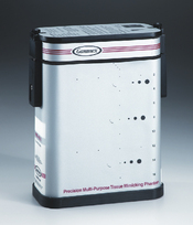
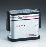
Commercially available test phantoms are normally filled with an aqueous gel or are formed from urethane materials. They contain various targets and structures designed for testing a number of parameters. Examples of some commercially available test phantoms are illustrated in Figure 13.1.


Fig. 13.1 Examples of some commercially available test phantoms used for ultrasound QA performance testing
(Reproduced with permission of Gammex.)

Stay updated, free articles. Join our Telegram channel

Full access? Get Clinical Tree