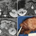
Fig. 1
ADK ductal adenocarcinoma
No: small NFET or other benign solid lesion
(?) B-mode:
Hypoechoic: ADK or NFET
Hyperechoic: focal fat accumulation
(?) CEUS
Hypoenhancing: ADK (→CT)
Hypervascular: NET (→CT/PET) or small solid SPT
Isovascular: MFP (→MRI)
(?) Liver metastasis
Yes: malignancy (→US-guided FNA)
(?) Imaging discordance:
Yes: EUS-guided FNA
Imaging Driving Questions in Cystic Pancreatic Lesions
General Imaging Findings
Some general findings can be assessed independently on the imaging technique.
(?) Shape
Cloud-like: SCA or BD-IPMN
Rounded: PSC or MCN or, less frequently, cystic NET, unilocular SCA or SPT
Oval and grape-like: IPMN
(?) Size
Very small: BD-IPMN or SCA
Small/intermediate: IPMN or SCA or PSC or MCN or cystic NET
Huge: PSC or MCA or cystic NFET or SPT
(?) Ductal dilation
Yes: MD-IPMN or MIX-IPMN or degenerated BD-IPMN
No: BD-IPMN or SCA or MCN
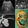
Ultrasound
As previously reported, US is very often the imaging method chosen as the first step for abdominal evaluation; therefore, also cystic pancreatic lesions may be identified. MRI with MRCP must always be performed to obtain a final diagnosis. EUS and EUS-guided FNA are valuable tools for cystic lesion characterization.
(?) B-mode:
Anechoic: IPMN, serous neoplasms
Inhomogeneous appearance: PSC, MCN, SPT, cystic NET
(?) CEUS
Enhancing areas:
Centrally oriented thin septa: SCA
Nodules, thick wall, and irregular septa: MCN
Nodules: degenerated IPMN
Inhomogeneous solid content: SPT
Thick wall: cystic NET
Avascular: PSC
(?) Imaging discordance or doubtful cases: EUS-guided FNA
Computed Tomography
CT should not be used for the evaluation of cystic pancreatic neoplasms, despite that some useful additional information, for example, regarding the presence of calcifications, could be provided.
(?) Pre-contrast
Hypodense: SCA, IPMN
Hyperdense: SPT or MCNs
Calcifications: MCN (peripheral) or SCA (central) or PSC (pancreatic)
(?) Post contrast
Enhancing portions
Centrally oriented thin septa: SCA
Nodules, thick wall, and irregular septa: MCN
Nodules: degenerated IPMN
Inhomogeneous solid content: SPT
Thick wall: cystic NET
Avascular: PSC
(?) Imaging discordance or doubtful cases: EUS-guided FNA
Magnetic Resonance
MRI is the imaging modality of choice for the comprehensive evaluation of patients with cystic pancreatic neoplasm.
(?) Communication with ductal system
Yes: IPMN
No: PSC or other cystic neoplasm
(?) T1-weighted images:
Homogeneously hypointense: IPMN, SCARelated posts:

Stay updated, free articles. Join our Telegram channel

Full access? Get Clinical Tree