, Ahmad Ameri1 and Mona Malekzadeh2
(1)
Department of Clinical Oncology, Imam Hossein Educational Hospital, Shahid Beheshti University of Medical Sciences (SBMU), Shahid Madani Street, Tehran, Iran
(2)
Department of Radiotherapy and Oncology, Shohadaye Tajrish Educational Hospital Shahid Beheshti University of Medical Sciences (SBMU), Tehran, Iran
The orbit contains the globe and connective tissues including extraocular muscles, orbital fat, eyelids, eyelashes, lacrimal gland, and lacrimal draining system. Different parts of the globe (e.g., the lens, iris, conjunctiva, sclera, retina, ciliary body, cornea, and optic nerve) have different sensitivities to irradiation. Some structures like the optic nerve respond to radiation in a chronic manner (late-responding tissue), and some like the conjunctiva and eyelids are acute-responding tissues. Some parts of the mentioned structures are very sensitive, and some of them like the lens and sclera are very resistant to radiation. The orbital area may be irradiated as the treatment target or as an organ at risk in the vicinity of the target. Radiation-induced acute side effects in this area require prompt attention to prevent long-term issues like ophthalmitis, permanent dry eyes, and impaired visual acuity. In this chapter, we will focus on more sensitive acute-responding tissues.
Most of the acute side effects of orbital radiation therapy have a mild severity, and there is often no need for treatment break [1–10]. These acute side effects (e.g., dry eyes, excessive tearing, conjunctivitis, transient periorbital erythema, and edema) have been reported in 30–50% of patients based on volume of the area within the radiation field [2, 3, 10–13]. Based on animal studies, the acute side effects of orbital radiation therapy in decreasing frequency include conjunctivitis, blepharitis, keratoconjunctivitis sicca, keratitis, and ulcerative keratitis [14].
The orbit is a small area that compared with its size, consists of a collection of many different structures as mentioned above. Each orbital structure has varying sensitivity to radiation effects and different signs and symptoms will develop at different times during radiation therapy. Here, the acute radiation effects will be reviewed for different orbital structures separately in each section.
4.1 Blepharitis and Eyelid Dermatitis
The eyelid is a thin fold of skin and its response to radiation is more than skin at other sites. It also contains hair follicles in its free edge that are sensitive to radiation. Radiation blepharitis (eyelid edge inflammation), as a radiation dermatitis, begins with erythema followed by dry and moist desquamation (and rarely necrosis). Erythema takes approximately 2–4 weeks to develop after radiation exposure; it is usually transient and disappears rapidly. Moist desquamation of skin can develop after 5–6 weeks of radiation therapy (50–60 Gy in 1.8–2.2 Gy daily fractions). Healing is typically slow and may take up to 6–12 weeks [15, 16].
The following guidelines will provide comfort and healing of the periocular skin radiation-induced dermatitis and blepharitis [17]:
Washing the eyelids with lukewarm water and mild soap and gently drying at least once daily.
Applying an eyelid moisturizer, black tea soaks (soak tea bags in hot water, allow them to cool, lie down, and put them over the eyes).
Avoiding extremes of heat and cold, skin irritants, and any eyelid rubbing or scratching friction.
More severe dermatitis in eyelid skin may need a radiation break and should be treated like a skin burn using silver sulfadiazine ointment or similar medications.
4.2 Eyelash Loss
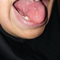
Loss of the eyelashes may occur (Fig. 4.1) with doses more than 10–20 Gy (1.5–2 Gy per fraction) [16, 18]. It may be temporary and regrowth after approximately 2 months [15] or may be permanent at radiation doses of more than 30 Gy [16].

Fig. 4.1
Conjunctivitis and eyelash loss at the end of radiation therapy for advanced maxillary sinus undifferentiated carcinoma
Eyelash loss can result in irritation of the conjunctiva and cornea due to loss of the protective blink reflex. These complications are discussed below.
4.3 Conjunctivitis
Signs and symptoms of radiation-induced conjunctivitis include conjunctival injection, watery discharge, and discomfort that occur 1–3 weeks after the start of radiation treatment. Acute conjunctivitis is common with doses ≥30 Gy [11, 19]. Secondary infectious conjunctivitis (mostly bacterial or rarely viral) may also occur [20] (Fig. 4.1). A thicker, purulent, yellow-green discharge should raise suspicion for bacterial conjunctivitis [21].
Radiation conjunctivitis is a clinical diagnosis based on patient signs and symptoms including a red eye, discharge, and normal vision as well as exclusion of other diagnoses in patients with a history of irradiation to or near the orbit [21].
The risk of conjunctival toxicity can be reduced by positioning the orbit out of the radiation field or reducing dose buildup on the conjunctival surface by higher beam energy or treating patients with the eye open during radiation exposure if eyes are irradiated from the anterior aspect of the orbit [20].
It is recommended that artificial tears be used 4–8 times daily to relieve the irritation caused by conjunctivitis [20].
Secondary infectious conjunctivitis should be managed as a primary form. Viral conjunctivitis is self-limited and topical antihistamine/decongestants may be needed for symptom relief. The efficacy of antiviral agents is not clear [22, 23]. Bacterial conjunctivitis is treated with a broad-spectrum topical antibiotic (trimethoprim with polymyxin B eye drops or erythromycin ophthalmic ointment). Gram stain or cultures generally are not carried out, and empiric antibiotics are usually prescribed [17, 24, 25].
4.4 Xerophthalmia
Tears are produced by the lacrimal functional unit consisting of the lacrimal glands, ocular surface (cornea and conjunctiva), eyelids, meibomian glands, and the interconnecting innervation [26, 27].
The tear film consists of three layers: mucous (inner layer), aqueous (middle layer), and lipid (outer layer).
The mucous layer is produced by conjunctival goblet cells and epithelial cells of the cornea and conjunctiva. The lacrimal glands consist of the main lacrimal gland and the accessory Krause and Wolfring lacrimal glands, which are located around the upper border of the tarsal plate and the conjunctival fornix, respectively. The aqueous component is secreted by these glands. The lipid component is secreted by the meibomian glands (at the rim of the eyelids inside the tarsal plate) and Zeis glands (anterior to the Meibomian gland, at distal eyelid margin) [28].
Radiation therapy leads to damage to the meibomian glands [29] and cause lacrimal gland acinar cell apoptosis and gland atrophy [30] or both [27]. Tear film aqueous and lipid deficiency develop due to dysfunction of lacrimal glands and Meibomian gland, respectively [20, 31].
With low-dose radiation therapy (about 24–25 Gy) of lymphoid lesions of the orbit and ocular adnexa, early mild radiation-induced xerophthalmia and chemosis have been noted in up to 50% of patients [12]. Irradiation of lacrimal glands to doses more than 30–40 Gy can lead to dry eye syndrome; the incidence increases dramatically with doses ≥50 Gy and doses greater than 60 Gy may cause permanent tear loss [20, 32, 33].
Diagnosis is based on characteristic symptoms and clinical appearance. Early effects are conjunctival inflammation, chemosis (swelling of the conjunctiva), and tear film instability with a resultant dry eye sensation [34]. Patients develop a red, painful, scratchy eye (foreign-body sensation), and photophobia. Severe problems may produce corneal desiccation, ulceration with bacterial infection, neovascularization, opacification, and ultimately perforation [35].
Xerophthalmia can be avoided by positioning lacrimal glands out of the radiation field, shielding them, or modifying dose distributions by using intensity-modulated radiation therapy (IMRT) [36, 37].
Supplemental lubrication or artificial tears are the mainstay of treatment for xerophthalmia; however, they act only as a replacement therapy without any effect on tear secretion. There are various artificial tear products with different formulations with no evidence suggesting a specific single brand being superior [38]. Preservative-free eye drops contain fewer additives and are generally recommended in the setting of extreme and long-term use of teardrop including severe dry eyes or multiple teardrop application daily due to lower effects of these products on the cornea and conjunctival epithelium [39].
In addition to artificial teardrops, there are other forms of supplemental lubrications like gels or ointments. Artificial tear ointments and gels offer longer-lasting relief but may blur vision [38].
There is no proven difference in efficacy between different topical dry eye treatments, including artificial tears, ointments, or gels [40].
Stimulation of tear production has been seen by using topical sodium hyaluronate, cyclosporine, and tacrolimus, which increase the aqueous component of the tear layer and goblet cell density while also decreasing the inflammation process [41, 42]. These agents need to be studied in radiation-induced xerophthalmia. Oral pilocarpine could palliate dry eye and dry mouth symptoms in Sjögren’s syndrome [43, 44] and has also been used in the management and prevention of radiation-induced xerostomia (see Chap. 7). But currently, there is no evidence for its application in radiation-induced xerophthalmia.
Topical anti-inflammatory agents (corticosteroids or nonsteroidal anti-inflammatory drugs (NSAIDs)) are beneficial for selective patients with severe dry eyes [45], but there isn’t sufficient evidence for their efficacy in radiation-induced xerophthalmia.
Autologous serum eye drops (with essential tear components for epithelial proliferation, similar properties of tears, and no additive preservatives) [46], punctal occlusion (mechanical blocking of the draining system) [47], and dry eye moisture chamber goggles (a chamber for protecting the dry eyes from the environment) [20], all have been suggested for severe dry eye treatment with some limitations in clinical use including need for frequent blood tests and potential risk of infection transmission in autologous serum eye drops, insufficient evidence of efficacy in punctal occlusion, and cosmetic concerns in wearing the moisture chamber goggles.
4.5 Corneal Toxicity
Acute corneal toxicity of radiation therapy can be secondary to radiation xerophthalmia or a primary effect of irradiation on the corneal surface epithelium, corneal stroma, and endothelium [20].
Secondary keratopathy due to tear film dysfunction is the common form of acute corneal toxicity and is presented by superficial punctate epithelial erosions or, in the case of severe dry eyes, corneal scarring [34].
Direct radiation injury can induce punctuate corneal erosions and corneal edema at doses of 40–50 Gy in 4–5 weeks and corneal ulceration at doses more than 60 Gy (with conventional fractionated radiation) and with 20 Gy delivered in a single dose [48].
Corneal damage is diagnosed clinically by signs and symptoms and eye examination including slit lamp exam. Patients usually present with photophobia, foreign-body sensation, tearing, pain, haloes, decreased vision, and red eye. In the penlight examination, conjunctival injection may be present. In slit lamp microscope examination, a corneal opacity or infiltration with corneal ulcers could be seen. The cornea may have a hazy appearance with corneal edema. Punctate epithelial erosions and corneal ulcers stain positively with fluorescein.
A corneal shield, which is placed between the lids and globe, could minimize corneal radiation dose and prevent toxicity if the treatment target is superficial to the cornea. These internal eye shields are used in radiation treatment planning of skin cancer near the eye and can give excellent protection from lower-energy X-rays and electron beams with energies ≥6 MeV [49, 50].
Related posts:

Stay updated, free articles. Join our Telegram channel

Full access? Get Clinical Tree


