Ill effects of radiation can be divided into two basic categories: stochastic effects and deterministic effects.1 Stochastic effects are those in which no clear relationship exists between the magnitude of the radiation dose and the severity of the effect. Stochastic effects include genetic mutation and induction of cancer. Estimations of the incidence of stochastic effects have been based on a no-threshold linear model assumption from the effects identified as a result of the atomic bomb detonations during World War II. These assumptions are not universally accepted. However, because of this uncertainty, the current approach is that stochastic effects have no threshold dose. Therefore, no radiation dose can be considered absolutely safe. It is imperative that fluoroscopically guided diagnostic and therapeutic procedures be performed under the safest conditions possible. Since the mid-1980s, skin injuries have been reported in patients as a direct result of complex fluoroscopically guided interventional procedures, including arterial embolization, arterial revascularization, cardiac radiofrequency ablation, and TIPS.2 The rise in reporting these adverse events resulted in U.S. Food and Drug Administration (FDA) action in 19943 and in U.S. federal regulations limiting the x-ray tube output of interventional fluoroscopic equipment. Similar actions have taken place throughout the world. The threshold dose for acute skin erythema is about 2 Gy, and that for delayed deep skin ulcers is about 12 to 15 Gy. The risk for deterministic injury rises if multiple sequential procedures are performed at the same anatomic location (i.e., a TIPS procedure and TIPS revision 3 months apart). Other risk factors for skin injury include obesity, diabetes mellitus, and connective tissue disease.4 Minimizing the risk of deterministic patient injury is a major focus of current radiation safety initiatives. The interventional procedure must be medically necessary to justify exposure to ionizing radiation. This is particularly important for procedures known to be associated with high exposure: TIPS, visceral stent placement, visceral embolization, and neuroembolization.5 For these interventions in particular, the risk for radiation-induced skin injury should be specifically discussed in the consent process. In addition, history taking should include questions about previous radiation exposure and factors that increase a person’s susceptibility to radiation-related skin injury (e.g., diabetes). The physical examination should include inspection of the skin at previous x-ray beam entry sites. Work habits of the interventionalist have a profound effect on patient radiation exposure.6 Optimal work habits require knowledge of radiation physics, familiarity with tableside controls of the x-ray equipment, practice using these controls, and a commitment to radiation safety as a patient care priority.
Radiation Safety and Protection in the Interventional Fluoroscopy Environment
Negative Effects of Ionizing Radiation
Management of Patient Exposure
Equipment Purchase and Maintenance
Preprocedural Patient Care
Intraprocedural Patient Care
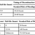
![]()
Stay updated, free articles. Join our Telegram channel

Full access? Get Clinical Tree


Radiation Safety and Protection in the Interventional Fluoroscopy Environment