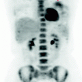
Fig. 14.1
MIP image: presence of slight and widespread increase in the glucose consumption of the axial skeleton, typical of bone marrow rebound
Modest increase in metabolism in the lower rectum-anus due to post-actinic fibrosis, SUV max 2.3.
14.4 Conclusions
The PET scan is negative for recurrence of disease with high glucose metabolism. (See Figs. 14.2, 14.3).
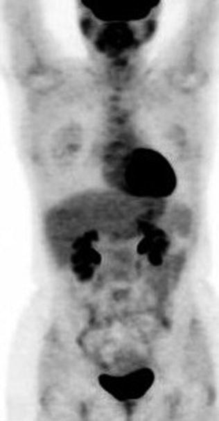
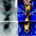
Fig. 14.2
 Laryngeal Squamous Carcinoma: Staging
 Sigmoid Adenocarcinoma: Post-Actinic Tardive Sacral Fracture
 Urothelial Carcinoma: Follow-Up After Surgery
 Lymphocytic Interstitial Pneumonia in Patient with History of Breast Cancer
 Metastatic Breast Carcinoma: Restaging After Neoadjuvant Chemotherapy
 Bone Metastases from Breast Cancer: Progression of Disease and Subsequent Response to Radiotherapy


Absence of lytic bone lesions on CT and focal areas with high metabolism at PET, excludes progression of skeletal disease
Related posts:
Laryngeal Squamous Carcinoma: Staging
Sigmoid Adenocarcinoma: Post-Actinic Tardive Sacral Fracture
Urothelial Carcinoma: Follow-Up After Surgery
Lymphocytic Interstitial Pneumonia in Patient with History of Breast Cancer
Metastatic Breast Carcinoma: Restaging After Neoadjuvant Chemotherapy
Bone Metastases from Breast Cancer: Progression of Disease and Subsequent Response to Radiotherapy
Stay updated, free articles. Join our Telegram channel
Full access? Get Clinical Tree
