4 Radiofrequency (RF) Coils
The closer the receiving antenna (or coil) is to the source of the MR signal, in general, the better the signal-to-noise ratio (SNR). As a natural consequence, an impressive variety of RF coils are available today, all designed to match the geometry and to allow optimal coverage of the anatomic region being examined. There are, however, important differences in internal design, the topic of this case, which are often not readily apparent on visual inspection. Also, although only rigid coils are illustrated, many modern coils (even the most advanced in design) are flexible, made so they can be wrapped closer to the body part of interest.
 Linearly Polarized Coils
The simplest coil, although rare today, is a single conductive loop (Fig. 4.1). The sum of the magnetic moments (the bulk magnetization) as it rotates causes a magnetic field fluctuation inside the loop area. This, according to Maxwell’s law, induces a voltage. The latter is digitized and analyzed to provide the information necessary to create an image.
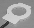
Fig. 4.1
 Circularly Polarized (CP) Coils
Stay updated, free articles. Join our Telegram channel

Full access? Get Clinical Tree


