Presentation and Presenting Images
( ▶ Fig. 4.1, ▶ Fig. 4.2, ▶ Fig. 4.3, ▶ Fig. 4.4, ▶ Fig. 4.5, ▶ Fig. 4.6, ▶ Fig. 4.7, ▶ Fig. 4.8)
A 72-year-old female with a history of invasive ductal carcinoma of the right breast treated with segmentectomy followed by oncoplastic tissue rearrangement and left breast mastopexy presents for asymptomatic screening mammography.
4.2 Key Images
( ▶ Fig. 4.9, ▶ Fig. 4.10, ▶ Fig. 4.11, ▶ Fig. 4.12, ▶ Fig. 4.13)
4.2.1 Breast Tissue Density
The breasts are heterogeneously dense, which may obscure small masses.
4.2.2 Imaging Findings
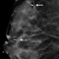
The imaging of the right breast demonstrates a postsurgical scar with architectural distortion and postsurgical clips (arrows in ▶ Fig. 4.9, ▶ Fig. 4.10, and ▶ Fig. 4.11) in the central aspect of the right breast middle and posterior depths. The imaging of both breasts demonstrates postsurgical changes consistent with mastopexy. In the right subareolar region there are radiolucent lesions with linear and curvilinear calcifications (circles).
The right craniocaudal (CC) tomosynthesis movie demonstrates the radiolucent lesions and calcifications on slices 17 to 52 of 90. A representative slice is shown (slice 24 of 90 in ▶ Fig. 4.12). Similarly, the area is seen on the right mediolateral oblique (MLO) tomosynthesis movie on slices 20 to 52 of 92. A representative slice is shown (slice 41 of 92 in ▶ Fig. 4.13).
4.3 BI-RADS Classification and Action
Category 2: Benign
4.4 Differential Diagnosis
Fat necrosis: The radiolucent lesions (fat) associated with linear and curvilinear calcifications are pathognomonic of fat necrosis.
Ductal carcinoma in situ (DCIS): While linear calcifications may be a presentation of DCIS, curvilinear calcifications are not.
Fibrocystic changes: Fibrocystic changes usually present as amorphous calcifications, not linear or curvilinear calcifications.
4.5 Essential Facts
The postsurgical history is important in this case. The patient has had right segmentectomy followed by right oncoplastic tissue rearrangement and left breast mastopexy. The right breast surgeries removed the breast malignancy and reestablished cosmesis. The left breast surgery established cosmesis. This history explains the appearance of both breasts.
There are many ways that fat necrosis may present mammographically, including lipid cysts, calcifications, focal asymmetries, or spiculated masses. If minimal fibrosis occurs, as in this case, then the mass appears radiolucent or as an oil cyst.
Oil cysts have a predictable evolution with linear and curvilinear calcifications developing and central calcifications developing later.
4.6 Management and Digital Breast Tomosynthesis Principles
Tomosynthesis, by removing overlapping tissue, easily delineates the radiolucent oil cysts and clearly demonstrates the associated calcifications along the edge of the oil cysts.
Even when unrecognized on conventional mammography, fat is commonly seen in both benign and malignant masses on tomosynthesis.
With tomosynthesis, the radiologist can confidently classify encapsulated fat-containing masses as benign.
Alternatively, radiologists must be careful not to misclassify all fat-containing lesions as benign or probably benign. Due to the growth of cancer, it can encapsulate fat and on tomosynthesis appear to contain fat. Fat-containing lesions should be carefully assessed for margins and other morphological features that could suggest a more suspicious lesion.
4.7 Further Reading
[1] Freer PE, Wang JL, Rafferty EA. Digital breast tomosynthesis in the analysis of fat-containing lesions. Radiographics. 2014; 34(2): 343‐358 PubMed
[2] Taboada JL, Stephens TW, Krishnamurthy S, Brandt KR, Whitman GJ. The many faces of fat necrosis in the breast. AJR Am J Roentgenol. 2009; 192(3): 815‐825 PubMed

Fig. 4.1 Right craniocaudal (RCC) mammogram.
Stay updated, free articles. Join our Telegram channel

Full access? Get Clinical Tree


