MR imaging plays a crucial role in the post-CRT assessment of rectal cancer; results are used for treatment planning. Radiologists should assess response or progression, possibility of a complete response, risk factors for incomplete resection, and nodal stage. T2-weighted MR imaging with diffusion-weighted imaging yields the best results to identify a complete response, but endoscopy is also very important. Overstaging of transmural and MRF invasion after CRT occur regularly, owing to residual stranding regarded as tumor to err on the safe side. Nodal restaging is a challenge. A structured report format or checklist is recommended.
Key points
- •
Restaging after chemoradiation in rectal cancer is crucial for treatment planning.
- •
Main challenges are identification of a complete response, extent of residual tumor and nodal stage after CRT.
- •
Complete response detection is best performed by combining T2-weighted MR imaging, diffusion-weighted imaging, and endoscopy.
- •
Use of a structured report or checklist is recommended.
Introduction
Many patients with rectal cancer are treated with neoadjuvant chemoradiation. Generally, in the United States, patients who have cT3 to 4N0 or any TN+ disease are candidates for neoadjuvant chemoradiation. Proximal T3N0 tumors sometimes may proceed to immediate resection; however, nodal metastasis is present in up to 22% of these patients, possibly leading to undertreatment if chemoradiation is omitted. , The purpose of this neoadjuvant treatment is to reduce tumor volume and stage to facilitate resection and to decrease local recurrence rates. Chemoradiation usually consists of a 5-FU-based agent (eg, capecitabine) and is combined with 45.0 to 50.4 Gy of radiation in daily fractions on weekdays. Restaging of the tumor after chemoradiation is performed around 8 to 10 weeks after the last radiation dose. Downsizing occurs in the majority of patients and even complete disappearance of the tumor and involved nodes occurs in approximately 16% of the patients (ypT0N0, complete response). The purpose of restaging is to reassess resectability of the tumor and to determine whether surgical treatment can be potentially avoided in patients with a complete response. In this article, the restaging of rectal cancer after chemoradiation is discussed, in particular the role of MR imaging.
Normal anatomy and imaging techniques
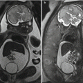
After chemoradiation, the main anatomy of the rectum and mesorectum remains the same; however, changes in the rectal wall, mesorectum, and surrounding structures can be seen. Owing to the irradiation submucosal edema occurs, which makes the rectal wall layer appear thickened and 3-layered on MR imaging (contrasting to a 2-layered wall in the normal untreated rectum; Fig. 1 ). This is usually most prominently visible in the distal rectum. Within the mesorectum, fluid and edema can be seen as well and fluid in the peritoneal reflection or along the mesorectal fascia is not an uncommon finding. Lymph nodes decrease in size (regardless of their metastatic status) and some disappear.

Restaging of rectal cancer after chemoradiation is mostly performed with MR imaging, but endoscopic ultrasound imaging can be used as an adjunct, to assess whether there is still a yT1 or yT2 tumor, if local excision is considered. MR imaging has the advantage of depicting the whole rectum and mesorectum, including surrounding organs and nodes, whereas with endoscopic ultrasound imaging these structures can only partially be evaluated by the limited field of view (just as in primary staging of rectal cancer). Endoscopy is an important additional tool to assess response and specifically when an organ preserving strategy is considered, endoscopy is crucial to perform next to MR imaging.
Imaging protocol
For restaging after CRT, the same protocol can be used as for primary staging, consisting of T2-weighted sequences in 3 orthogonal directions (sagittal, coronal, and axial), where the axial plane is perpendicular to the (former) tumor axis and the coronal plane is parallel. An axial DWI (exact same angle as the axial T2-weighted sequence) is important in restaging after CRT (including a high b-value of 800–1000). The apparent diffusion coefficient (ADC) map must be constructed and send to the PACS. An unenhanced T1-weighted large field of view sequence can be considered to depict the whole regional lymph drainage route and it can be helpful to assess bony changes in some cases. If a large field of view T1 sequence is not used, the T2 sequence should be scanned with a large field of view. It is important to compare the post-CRT images with the pre-CRT MR imaging during MR imaging acquisition to identify the (former) tumor location, because, owing to tumor response, this can be challenging sometimes. For restaging purposes use of an enema before the MR imaging has been shown to significantly decrease artifacts, which make the evaluation of DWI easier; therefore, the use of an enema is recommended. Ultrasound gel shows T2 shine through and is not recommended, because it obscures any residual high signal of tumor in the rectal wall.
Table 1 shows an example of an MR imaging protocol, as used in the authors’ institution.
| Sagittal T2 SE | Axial T2 SE | Coronal T2 SE | T1W | DWI | |
|---|---|---|---|---|---|
| TR | 3357 | 11,838 | 8456 | 9.8 | 2829 |
| TE | 150 | 130 | 130 | 4.6 | 95 |
| NSA | 3 | 2 | 2 | 1 | 8 |
| Acquired in plane resolution (mm) | 0.78 × 1.12 | 0.78 × 1.15 | 0.78 × 1.15 | 1.14 × 1.15 | 2.25 × 2.32 |
| Matrix | 256 × 178 | 256 × 174 | 256 × 174 | 264 × 261 | 80 × 65 |
| Slice thickness (mm) | 4 | 3 | 3 | 1 | 5 |
| Slices | 22 | 42 | 30 | 200 | 20 |
| Time (min) | 4:43 | 5:43 | 4:05 | 6:11 | 1:33 |
| EPI factor | 65 | ||||
| b-values | 0, 1000 | ||||
| Fat suppression | SPIR |
Imaging findings and diagnostic criteria
Most of the tumors will respond to chemoradiation and show a degree of response. For treatment purposes it is important to answer the following questions:
- •
Is there residual luminal tumor?
- •
If these is residual tumor, what is the ycT stage?
- •
Are there any findings that indicate a risk for incomplete resection?
- •
Is there (residual) nodal involvement?
Is There Residual Luminal Tumor?
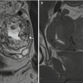
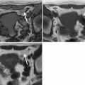
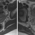
As mentioned, many tumors show response. When a tumor responds to chemoradiation, tumor tissue is replaced by fibrosis. Fibrosis has a hypointense signal. The distribution of the fibrosis tends to follow the distribution of the tumor: that is, if the tumor was circular, the fibrosis will also be circular. Also, the shape will generally not change, for example, an irregular spiculated tumor will show irregular fibrosis as well. A complete response (ypT0) will show either a normalized rectal wall (rare, approximately 5% of cases with ypT0) or homogeneous fibrosis without diffusion restriction. However, irregular more heterogeneous fibrosis or fibrosis with focal diffusion restriction sometimes do correspond with a complete response after surgery. The sensitivity of T2-weighted imaging for the prediction of a complete response is low (19%). Patel and colleagues recommended to use the mr tumour regression grade (mrTRG) system, which is a radiologic counterpart for the TRG system that is used in histopathology to grade response. Essentially, the mrTRG is a method to assess the amount of fibrosis and residual tumor, which is very similar to but more standardized than standard evaluation of the response to CRT. Unfortunately, the sensitivity to detect a complete response is still only 74% when using mrTRG. The addition of a DWI sequence increases overall diagnostic performance to 80%, when using the presence of absence of diffusion restriction as a diagnostic criterion. In these cases, sensitivity was reported to be rather disappointing (52%–64%), meaning that many complete responses are missed. A more pattern-based approach has been proposed to improve diagnostic performance. Similar to the morphologic response at T2-weighted (TW2) MR imaging, response on diffusion tends to follow the shape of the tumor as well. Polypoid or semicircular tumors usually show focal diffusion restriction, whereas circular tumors have scattered areas of diffusion restriction or circular more massive diffusion restriction. By use of such a pattern-based approach, a sensitivity of 94% and specificity of 77% could be reached to predict complete response in experienced hands. The results of this study are promising and can hopefully be reproduced in a prospective setting. Fig. 2 shows an example of a complete response on T2W MR imaging and DWI.


