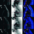
Fig. 30.1
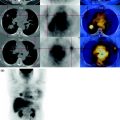
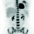
The MIP image shows the extent of the disease in the pelvis and the intensity of the metabolic activity of the numerous metastatic masses. In this reconstruction, the pulmonary nodule is not seen because the limited metabolism of the lesion is masked by the physiological activity of the mammary glands
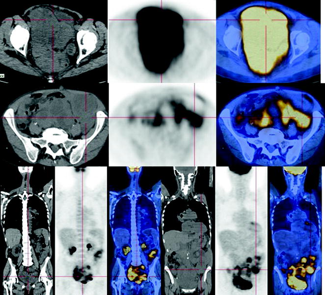
Fig. 30.2


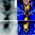
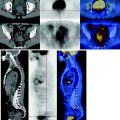
CT shows the voluminous homogeneous, partly liquid and solid, widely confluent and necrotic masses occupying the pelvis. The coronal reconstruction shows the extension of the upper abdominal disease, especially to the left. The PET shows increased consumption of glucose with contextual areas with low metabolism contained within, due to the presence of liquid and or colliquative component
Stay updated, free articles. Join our Telegram channel

Full access? Get Clinical Tree


