Relapsing Polychondritis
Aqeel A. Chowdhry, MD
Tan-Lucien H. Mohammed, MD, FCCP
Key Facts
Terminology
Rare autoimmune episodic disorder that destroys cartilage, especially of ear, nose, and laryngotracheobronchial tree
Imaging Findings
Malacia earliest finding, probably secondary to edema and cartilage inflammation
Airway wall then becomes thickened and cartilaginous portions start to calcify
Stenosis late finding with scarring
Increased attenuation of airway wall on noncontrast CT classic, up to 100%
Spares posterior tracheal membrane (no cartilage)
Calcification is smudgy or putty-like
Top Differential Diagnoses
Wegener Granulomatosis
Tracheopathia Osteochondroplastica
Amyloidosis
Laryngotracheal Papillomatosis
Clinical Issues
Prolonged remitting disease, diagnosis usually delayed 3 years
Swelling and redness of ears (90%)
Nasal chondritis (50%) results in saddle nose deformity
Respiratory complications account for 30% of deaths
Hearing loss (50%), often sudden
Diagnostic Checklist
Sparing of posterior membrane tracheal wall key to differentiate from other causes of diffuse tracheal disease
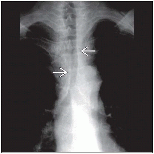 Frontal radiograph shows diffuse thickening of the tracheal wall  in a patient with relapsing polychondritis. Tracheal pathology is often overlooked on chest radiography. in a patient with relapsing polychondritis. Tracheal pathology is often overlooked on chest radiography. |
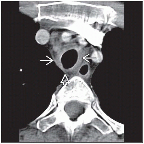 Axial CECT shows smooth thickening of the trachea  . The airway wall is slightly increased in attenuation, but the posterior trachea . The airway wall is slightly increased in attenuation, but the posterior trachea  is normal. is normal. |
TERMINOLOGY
Definitions
Rare autoimmune episodic disorder that destroys cartilage, especially of ear, nose, and laryngotracheobronchial tree
IMAGING FINDINGS
General Features
Best diagnostic clue: Increased thickness and attenuation of tracheal wall
Patient position/location: Trachea and main bronchi most common
Morphology: Diffuse thickening of tracheal wall, sparing of posterior tracheal membrane
CT Findings
Central airways
Airway wall thickening
Distribution: Trachea & bronchi > trachea only > bronchi only
Diffuse with smooth external and internal contours (90%)
Focal and nodular uncommon
Increased attenuation of airway wall (100%)
Ranges from subtle increase in attenuation to calcification
Calcification is often smudgy or putty-like
Involves cartilaginous portions of airway only
Dimensions
Diffuse narrowing (50%)
Focal (50%), usually subglottic location
Distribution
Involves cartilaginous airway wall only
Spares posterior tracheal membrane (no cartilage)
Evolution
Malacia earliest finding, probably secondary to edema and cartilage inflammation
Airway wall then becomes thickened, and cartilaginous portions start to calcify
Stenosis late finding with scarring
Lung
Air-trapping and bronchomalacia (50%)
Lobular air-trapping > segmental air-trapping > lobar air-trapping
Mild cylindrical bronchiectasis (25%) due either to recurrent infections or direct destruction of cartilage
Diffuse alveolar hemorrhage associated with glomerulonephritis
Cardiovascular
Aneurysmal dilatation of aorta, especially ascending aorta root
Aortic wall thickening if aortitis
Cardiac enlargement: Aortic or mitral valve regurgitation or from pericarditis
Radiographic Findings
Radiography: Trachea common blind spot in radiography; pathology often overlookedRelated posts:

Stay updated, free articles. Join our Telegram channel

Full access? Get Clinical Tree


