AAST classification
Description of injury
Grade I
Contusion or non-expanding subcapsular hematoma with no laceration. Constitutes 80 % of patients
Grade II
Non-expanding perirenal hematoma confined to the retroperitoneum
Laceration <1 cm deep without collecting system injury and urinary extravasation
Grade III
Cortical laceration >1 cm without collecting system injury
Grade IV
Laceration: through corticomedullary junction into collecting system
Vascular: involves main renal artery or vein with contained hematoma, segmental infarctions
Grade V
Laceration: shattered kidney, ureteropelvic junction avulsion
Vascular: renal pedicle injury or avulsion, devascularization of the kidney

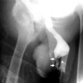
Fig. 9.1
The AAST grading of renal injury. (a) Grade I, subcapsular hematoma (arrow). (b) Grade II, cortical laceration less than 1 cm deep (arrow) and perinephric hematoma (arrowhead). (c) Grade III, cortical laceration more than 1 cm deep (arrows) and perinephric hematoma. (d) Grade IV, laceration extending through the renal cortex, medulla (arrows), and collecting system (curved arrow). (e) Grade IV, segmental infarction caused by thrombosis of segmental renal artery branches (arrows). (f) Grade V, shattered kidney. (g) Grade V, avulsion of renal hilum that devascularizes the kidney (With permission from Lee YJ, Oh SN, Rha SE, Byun JY. Renal trauma. Radiol Clin North Am. 2007;45:581–92)
Shattered kidney refers to extensive fragmentation of the kidney and is usually associated with varying degrees of renal parenchyma devitalization (Fig. 9.2a–d).
Fig. 9.2
Shattered kidney (grade V) with ureteropelvic junction laceration in a 30-year-old man who had an underlying horseshoe kidney. (a) Contrast-enhanced axial CT scan in the nephrographic phase shows multiple fragments of renal parenchyma (arrows) at the fusion site of the two kidneys and a large amount of hematoma in the anterior and posterior pararenal spaces, bilaterally. (b) Contrast-enhanced axial CT scan shows extravasation of contrast media (arrows). (c) Multiplanar reformatted image in the coronal plane in the nephrographic phase shows a shattered horseshoe kidney with a large amount of hematoma in the perinephric space (arrows). (d) In a volume-rendered oblique coronal image, a large volume of contrast media extravasation (arrows) from the left kidney is readily apparent (With permission from Lee YJ, Oh SN, Rha SE, Byun JY. Renal trauma. Radiol Clin North Am. 2007;45:581–92)
Renal injury caused by trauma is usually treated conservatively except in cases of severe injury such as pedicle injury or complete laceration of the ureteropelvic junction.
Box 9.1 Indications for Renal Imaging After Trauma
Gross hematuria
Rapid deceleration injury
Blunt trauma and shock with gross or microscopic hematuria
Imaging
Plain Film Radiography
Secondary findings such as rib fractures, asymmetry of the soft tissues overlying the flanks, and asymmetry of the psoas shadows may suggest possible renal injury.
Intravenous Pyelography
IVP study consists of scout radiograph, one film immediately after contrast injection, and another 10 min after contrast injection.
Blurring of the renal outline or psoas shadow due to perirenal hemorrhage may be seen on IVP.
An injured kidney may exhibit delayed, diminished, or absent excretion of contrast material.
Nonvisualization, contour deformity, or extravasation of contrast medium on IVP indicates a major renal injury.
The use of IVP to evaluate renal trauma has been largely supplanted by CT scan, which is the preferred modality for this type of study.
Ultrasound
Ultrasound detects about 22 % of renal injuries.
Ultrasound may reveal renal lacerations, changes in echogenicity of renal parenchyma and/or renal pelvis, and decreased echogenicity of perirenal space.
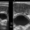
Ultrasound reveals free or loculated fluid around the kidney with variable appearance depending on the type of fluid (urine or hemorrhage) (Fig. 9.3).
Fig. 9.3
Renal subcapsular hematoma secondary to iatrogenic trauma. Sagittal view on gray-scale ultrasound demonstrates subcapsular hematoma (arrows) secondary to ESWL with hypoechoic appearance
FAST scan (focused abdominal sonography for trauma scan) is mainly useful in detecting free fluid in the abdomen.
Negative ultrasound findings do not exclude the possibility of renal injury.
Computed Tomography
CT can assess the extent and spatial location of traumatic renal injuries.
Parenchymal contusions appear on CT as round or ovoid areas of decreased enhancement with ill-defined contours (Fig. 9.4a, b).
Fig. 9.4
Renal contusion. (a) Axial and coronal (b) views of contrast-enhanced CT demonstrate ill-defined hypodense renal parenchymal contusion (arrows) in the left kidney. Retroperitoneal hematoma (*) secondary to trauma is also observed
Parenchymal lacerations manifest on CT as hypodense linear or wedge-shaped parenchymal defects or clefts (Fig. 9.5a, b). If lacerations contain blood clot, they may appear as hyperdense parenchymal clefts.