Renal Artery Embolization
Clinical Relevance
Embolotherapy of the renal arteries was introduced into the portfolio of the interventional radiologist many years ago. It was most popular around 25 years ago when preoperative embolization of kidneys containing a renal cell carcinoma (hypernephroma) was widely used as a routine procedure in the belief that preoperative embolization might avoid tumor seeding during surgery. Since no oncological benefit has since been proven by this strategy, and blood loss is no longer a major problem in renal surgery, preoperative embolization is now limited to a few uncommon circumstances. Nevertheless, many different techniques were developed, resulting in the current array of indications for renal embolotherapy.
Indications
Tumor Embolization
Total renal embolization is indicated for inoperable hypernephromas in patients with persistent hematuria or severe paraneoplastic symptoms such as hypercalcemia (Stauffer syndrome). In these patients, complete embolization of the tumor-bearing kidney is indicated if a functioning contralateral kidney is still present. Some groups prefer tumor embolization even without symptoms to reduce tumor bulk in combination with immunostimulating therapies. In total embolization not followed by nephrectomy, embolization should be performed by particles of small diameters or bucrylate–ethiodized oil mixed in a ratio of 1 : 3 to 1 : 5 to allow deep deposition into the tumor tissue.
Preoperative embolization is still indicated in a small subset of tumors, especially those that have developed extensive venous involvement. Preoperative embolization may be indicated as a safety procedure in patients who refuse blood transfusions, such as Jehovah’s witnesses. In preoperative embolization, the technique is modified because only occlusion of the main renal artery is necessary. This can be achieved by coils, Amplatzer plugs, or bucrylate–ethiodized oil mixed in a ratio of 1 : 1 to 1 : 3 to achieve rapid occlusion of the main renal artery.
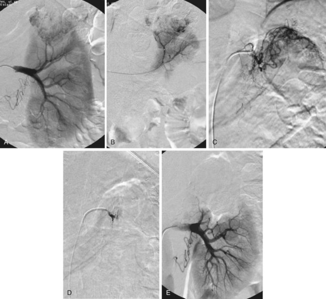
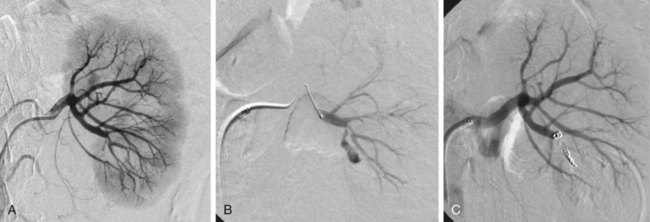
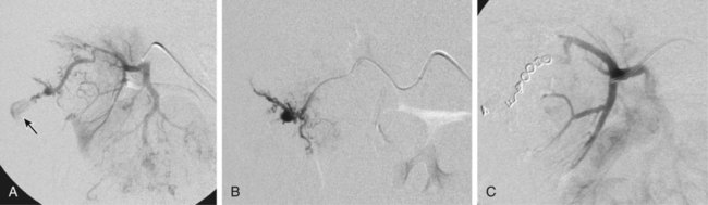
Partial tumor ablation by embolization (Fig. 60-1) is preferred in patients who have a tumor in a single functioning kidney. Partial embolization has also been used as a preparation for thermal ablation of a circumscribed peripheral malignant tumor to reduce the risk of postprocedural bleeding after radiofrequency ablation or to enhance its efficacy.1

There is more published experience of renal artery embolization for ruptured or bleeding benign renal tumors such as angiomas and particularly angiomyolipomas.2 The latter have a tendency to grow during pregnancy, and prophylactic embolization may be performed in women who wish to become pregnant to prevent major bleeding during pregnancy. In benign lesions, a partial embolization is preferable whenever possible.
Defunctionalization
Total renal embolization, including bilateral embolization in some cases, may be indicated in patients with massive protein loss in nephrotic syndrome or other complications of end-stage renal failure such as intractable hypertension. Other indications for total renal embolization are failing kidney transplants, as an alternative to surgical removal in cases of graft intolerance syndrome, and persistent urinary leaks in failing kidneys.3
Very rarely, partial defunctionalization has been described in cases with segmental arterial stenosis and hypertension where the responsible segmental artery was embolized.4 Alternatively, these patients may undergo intrarenal percutaneous transluminal angioplasty (PTA) using small balloons.
Bleeding Control
Blunt or direct trauma may cause renal bleeding due to renal laceration or renal artery rupture. Trauma may also cause renal artery pseudoaneurysms, arteriovenous (AV) fistulas combined with hematuria, direct hemorrhage into the pelvicalyceal system, or development of perirenal hematomas.
Unfortunately, the main causes of traumatic renal hemorrhage are iatrogenic in origin and represent the majority of cases of traumatic renal hemorrhage in Europe. They include renal biopsies in native and transplant organs, percutaneous techniques such as percutaneous nephrostomy (Fig. 60-2), nephrolithectomy, and percutaneous transluminal renal angioplasty (PTRA), where direct arterial perforation by the guidewire tip has been described.5 Also, indirect methods such as shockwave lithotripsy can lead to renal trauma requiring embolotherapy.

Although iatrogenic hemorrhage is relatively common, few cases require treatment (0.3%-1% of percutaneous renal interventions6 and 0.5% of percutaneous biopsies7). Moreover, although hemorrhage that requires treatment is unusual, temporary and self-limiting bleeding or AV fistulas occur in up to one third of percutaneous renal procedures.7 Since the introduction of partial nephrectomy techniques, bleeding after partial surgery has become a significant cause of iatrogenic hemorrhage. Approximately 2% of patients who undergo partial nephrectomy develop postsurgical bleeding, and most of them require percutaneous embolization.8
Rarely, benign tumors such as angiodysplasias, angiomas, or angiomyolipoma (Fig. 60-3) may cause spontaneous—sometimes substantial—bleeding (Wunderlich syndrome)9 and may undergo embolization. In a few cases, they may also develop hypertension.

Renal Artery Aneurysms
As a special problem, true and false aneurysms of the mainstem renal artery10 may occur that may be prone to rupture or serve as an embolic source for renal infarction. There is a general consensus that a diameter of 20 mm is a threshold for treatment; smaller aneurysms may undergo treatment if severe hypertension is present and difficult to treat. Central renal artery aneurysms require treatment by coil placement or, if possible, exclusion by the use of stent grafts. Peripheral (pseudo)aneurysms may be due to trauma or systemic diseases such as polyarteritis nodosa. Peripheral aneurysms are best treated by exclusion, coil placement, or medical adhesive (cyanoacrylate, “glue”) deposition. The most difficult location for treatment of renal artery aneurysms is at the site of a renal artery bifurcation (or trifurcation), because exclusion of the aneurysm may require occlusion of one or more segmental arteries. A combination of bare metallic stents with coils, glue, Onyx (ethylene vinyl alcoholic copolymer [ev3/Covidien, Plymouth, Minn.]), or flow-diverting stents may be an option in these challenging locations.10–14
Contraindications
There are few absolute contraindications to total or selective renal artery embolization. Embolization should be avoided in the presence of acute infection to avoid superimposed infection of the devascularized territories.
Relative contraindications are impaired global renal function, allergic reactions to contrast media, acute hyperthyroidism, planned radioiodine therapy, and single kidneys. In the latter, embolization may be performed if no other nephron-sparing surgical alternative exists.
Technique
Anatomy and Approaches
Depending on the underlying disease, a complete or partial embolization is performed. Classical indications for complete renal embolization are embolotherapy before tumor nephrectomy, total renal ablation in tumors where there is a contraindication to surgery, or defunctionalization for benign causes either in native kidneys or transplants.
Partial embolization is performed to preserve renal function when it is possible to treat a localized lesion, and can be used in traumatic or iatrogenic lesions as well as in single kidneys with renal artery tumors. Whereas in total renal artery embolization, complete destruction of functioning renal tissue is the aim of treatment, when partial renal embolization is performed, the aim is to minimize loss of normal renal parenchyma.
Technical Aspects
Many materials have utility for renal arterial embolization. For total renal embolization, absolute ethanol is a common embolic agent used. Other liquid embolics such as glue (bucrylate [Histoacryl; Braun, Melsungen, Germany]) may be used after mixing with ethiodized oil (Ethiodol, Lipiodol [Guerbet USA, Bloomington, Ind.]). Total embolization with embolic microspheres (e.g., polyvinyl alcohol [PVA] particles), using a small particle size between 20 and 500 µm, has also been described.
In partial embolization, the choice of embolic agent depends on the purpose of treatment. In selective tumor embolization, the tumor bed is preferentially embolized using small particles after superselective catheterization of the tumor-feeding renal artery branches. Alternatively, embolization with glue mixed with ethiodized oil may be performed. If the main feeding branch can be identified, it can be additionally occluded by coil placement or a bucrylate–ethiodized oil mixture. If the patient is being treated for a vascular complication such as AV fistula, pseudoaneurysm, or blunt traumatic hemorrhage, a very localized embolization is desired, either by coil embolization or local application of glue (bucrylate–ethiodized oil).

Full access? Get Clinical Tree

