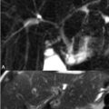
101 Renal Artery MRA Contrast-enhanced MRA is also commonly utilized for the noninvasive evaluation of renal artery stenosis—the most common cause of secondary hypertension. Figure 101.1 demonstrates focal high-grade stenosis of the right renal artery on (A) coronal contrast-enhanced MIP MRA images. Focal, less-severe narrowing at the left renal artery origin is also present. Typically, only renal artery stenoses greater than 50% respond to surgical therapy; however, significant drops in arterial blood flow may not occur until stenoses exceed 75% in degree. Some stenoses occur without associated parenchymal dysfunction as evidenced by the fact that not every patient with moderate- or high-grade stenosis responds to surgical treatment. Atherosclerotic causes of renal artery stenosis predominate, preferentially involving the proximal third of the renal artery and the ostium. Measurements of renal artery stenoses are conveniently performed on coronal source images but suffer intraobserver variability and assess the stenosis in only one dimension. Image acquisition with isometric voxels allows reconstruction in any desired plane, as illustrated by the (B) sagittal, showing the origins of the superior mesenteric and celiac arteries, and (C) axial MIP reconstructions, both obtained from the same coronal source image dataset as was the coronal MIP in Fig. 101.1A. A more reproducible approach to stenosis measurement thus involves initial identification of potential stenoses on coronal MIP images with subsequent measurements performed on reconstructions perpendicular to the region of suspected stenosis and also in the sagittal plane.
Stay updated, free articles. Join our Telegram channel

Full access? Get Clinical Tree


