Composition
Frequency (%)
Men
Women
Radiopaque stones
Calcium oxalate – calcium phosphate
92.1
90.3
Struvite
1.4
5.1
Radiolucent stones
Cystine
0.7
1.6
Uric acid
5.5
2.2
Others
0.3
0.7
With modern scanners and state of the art scanning protocols; asymptomatic, low density, and/or millimetric stones can be easily detected.
Urinary Stone types
(a)
Calcium oxalate stones: 70 % of all urinary stones contain calcium oxalate. Dietary factors, inborn errors of metabolism, and acquired metabolic disorders (disturbances of calcium metabolism) predispose to calcium oxalate stone formation. A high oxalate excretion is associated with high risk of recurrent stone formation. A low excretion of citrate has been demonstrated in up to 50 % of patients with calcium-oxalate disease.
(b)
Struvite stones (triple phosphate stones): Struvite stones are common in patients with urinary tract infection with pseudomonas, proteus, klebsiella, and streptococcus. These organisms produce urea-splitting enzymes, resulting in increased concentration of ammonia and bicarbonate in the urine, thereby predisposing patients to an increased incidence of struvite stones (infection stones). Struvite stones are composed of magnesium ammonium phosphate, calcium carbophosphate, carbonate apatite, and urease. Seventy percent of staghorn calculus are struvite stones.
(c)
Cystine stones: Cystine stones are more common in adults than in children. Cystinuria is an autosomal recessive disorder resulting in abnormal transport of cysteine, ornithine, lysine and arginine in the proximal renal tubule, and intestinal epithelium. Cystine solubility coefficient in urine is very low, resulting in stone formation.
(d)
Uric acid stones: Primary uric acid stones occur in Lesch-Nyhan syndrome, and secondary urate stones are seen in patients with inflammatory bowel disease, lymphoproliferative disorders, and myeloproliferative disorders.
(e)
Oxalate stones: These stones are associated with hyperoxaluria of two types: primary hyperoxaluria (Type I) is the result of an autosomal recessive disorder, in which the defect lies in alanine-glycolate aminotransferase causing hyperglycosuria. Type II hyperoxaluria is secondary to a defect in glyoxylate-reductase. The presence of glycolate in type I can be confirmed by liver biopsy and this is pathognomonic. Secondary type of oxalate stones occur in conditions such as cystic fibrosis, inflammatory genitourinary disorders, and ileal resection.
Lasix Stones
Lasix (furosemide) stones are common in neonates treated with Lasix for congestive heart failure. Lasix induces calciuresis and stimulates the parathyroid gland, causing secondary hyperparathyroidism. Lasix stones do not require any specific treatment as these stones disappear after cessation of the use of Lasix.
Proteinase Inhibitors and Urinary Stones
Indinavir and atazanavir are used in the treatment regimen for patients with AIDS. These drugs act as a protease inhibitors, ultimately preventing the formation of new viral particles. Nephrolithiasis induced by indinavir and atazanavir is reported with an incidence of approximately 12.4 and 7.3 %, respectively. Darunavir is a second generation protease inhibitor; the incidence of urinary stone formation in patients treated with darunavir is reportedly 0.15 %.
Imaging
Plain Film Radiography
The basic imaging modality for suspected urinary calculus disease is plain film radiography.
Kidney-ureter-bladder (KUB) can detect urinary stones that are radiopaque (Fig. 4.1).
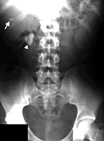
Fig. 4.1
Staghorn right renal pelvic stone (arrowhead) is seen on this KUB. In addition a gallstone (arrow) is present
Typically, urinary stones are found in the renal collecting system, the bladder, or at locations where the ureters are narrower, such as ureteropelvic junction, iliac vessel crossing, and ureterovesical junction (Fig. 4.2).
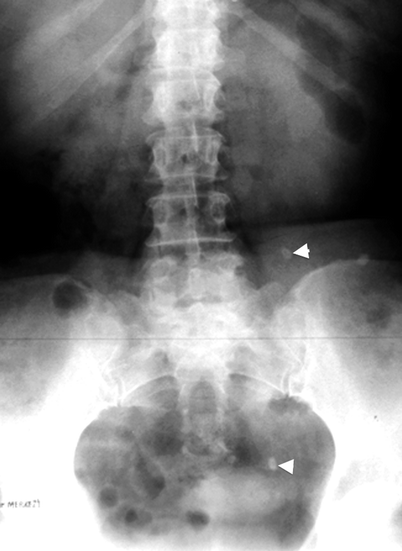
Fig. 4.2
KUB demonstrates radiopaque left ureteral stones (arrowheads) at middle and distal segments of the ureter
Radiopaque upper urinary tract stones are usually easy to define with their unique morphological features, such as staghorn shape or faceted contours.
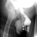
Renal stones that involve the renal pelvis and at least two calyces are classified as staghorn calculi. These stones are mostly composed of mixtures of magnesium ammonium phosphate and/or calcium carbonate apatite (Figs. 4.3 and 4.4a–c).
Fig. 4.3
The left pelvicalyceal system is completely filled with a large staghorn calculus
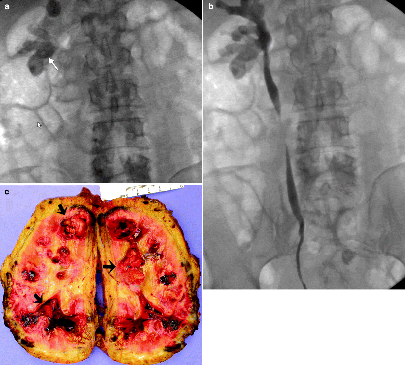
Fig. 4.4
Staghorn calculus. (a) Plain film radiograph (inverted image) demonstrates staghorn calculus (arrow) in the right kidney. (b) Retrograde pyelography reveals staghorn calculus involving the middle and lower pole calyces of the right kidney. (c) Nephrectomy specimen, bivalved, from a patient with recent sepsis and nonfunctioning kidney. Arrows indicate multiple soft calculi (struvite stones) in the renal pelvis and calyces
Renal outlines can often be clearly delineated, as kidneys are completely surrounded by retroperitoneal fatty tissue, so secondary findings such as renal enlargement and obscured renal margins related to obstructing stones, whether they are radiopaque or radiolucent, may also be detected by KUB examination.
Scoliosis with a concavity towards the painful side is another useful additional sign in detecting a renal stone and localizing its side.
KUB has several drawbacks:
Radiolucent stones cannot be detected.
Patient’s body habitus or bowel contents may mask small radiopacities.
Mid-ureteric stones are not easy to detect because of sacral superposition (Fig. 4.5).
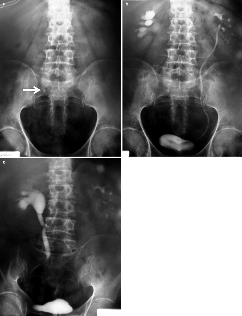
Fig. 4.5
Mid-ureteric stone. (a) On this KUB, millimetric right ureteric stone is superposed over sacrum (arrow). Fifteenth minutes (b) and late (prone position) (c) IVP series reveal delayed right renal function
Bowel preparation and oblique projections may facilitate the diagnosis when needed.
Urinary stones may be found at unexpected sites due to structural variations or anomalies of the urinary system, such as malrotation and ectopic location (Fig. 4.6).
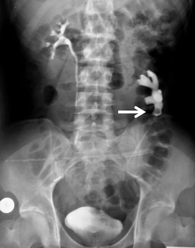
Fig. 4.6
Left lower calyx stone, which is seen as a filling defect on this IVP spot film (arrow), is located unexpectedly far away from midline secondary to the ectopic and mal-rotated left kidney
Distinction of stones from various extra-urinary radiopacities may be difficult.
Cholelithiasis, calcified nevi, or lymph nodes may mimic upper urinary tract stones, whereas phleboliths and calcifications related with internal genital organs and diseases may resemble lower urinary tract stones (Figs. 4.7, 4.8, 4.9, and 4.10).
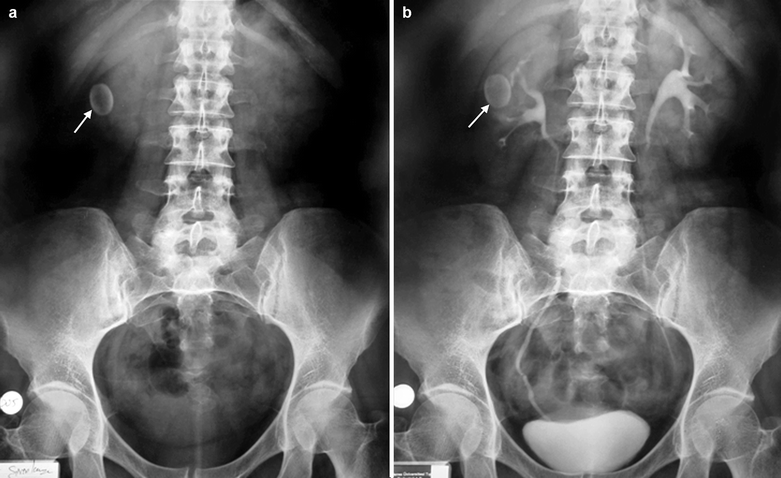
Fig. 4.7
Gallbladder stone. KUB and IVP spot films reveal an ovoid shaped gallbladder stone (arrows) superposed on the right kidney (a, b)
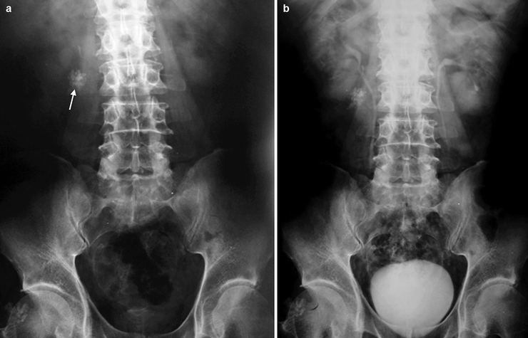
Fig. 4.8
Calcified nevus. (a) An amorphous calcification (arrow) is present at right paravertebral location on KUB. (b) IVP spot film shows that the lesion is unrelated to the urinary tract. On detailed inspection of the patient, a calcified nevus was seen on the right lumbar region
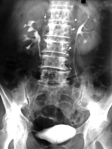
Fig. 4.9
Calcified lymph node. Bilateral and multiple calcified retroperitoneal lymph nodes are visible on IVP (arrows)
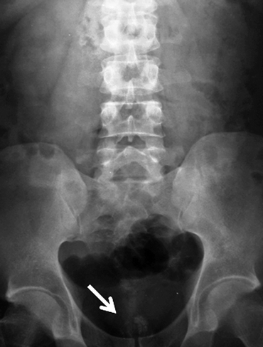
Fig. 4.10
Calcified corpora amylacae. Midline amorphous calcification cluster (arrow) is present at bony pelvis floor which is consistent with corpora amylacea of the prostate
Detailed inspection of the patient or additional imaging studies are occasionally needed to characterize the suspicious lesion exactly.
Intravenous Pyelography
The traditional imaging modality of the urinary system is IVP.
Comprehensive data about the functional and morphological characteristics of the urinary system can be obtained with a couple of projections after intravenous administration of water soluble and nonionic contrast media.
The technique is also the preferred imaging modality by urologists because minimally invasive interventions, such as extracorporeal shock wave lithotripsy and percutaneous nephrolithotomy, can be accurately planned according to collecting system anatomy revealed by IVP.
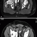
Radio-opaque stones are usually located by IVP but may be obscured by contrast material (Figs. 4.11, 4.12, and 4.13).
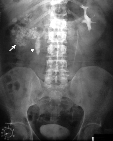
Fig. 4.11
Multiple millimetric radiopaque renal stones (arrow) and a large proximal ureter stone (arrowhead) are present on the right side. Right renal function is deteriorated
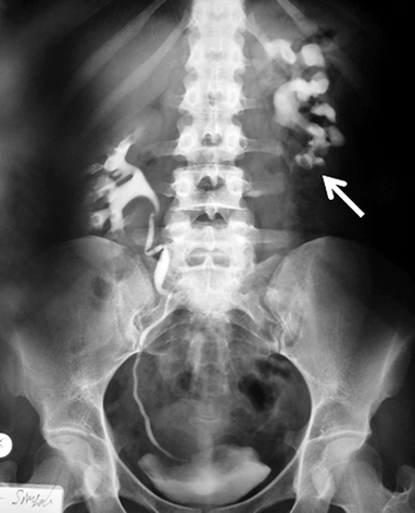
Fig. 4.12
Left lower pole calyx stone (arrow) is completely covered by contrast material (KUB not shown)
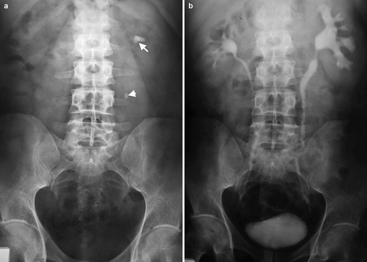
Fig. 4.13
Left uretero-pelvic (arrow) and mid-ureteric stones (arrowhead) are covered with contrast material on IVP series (a, b). Mild hydroureteronephrosis is seen secondary to these partially occluding stones
In contrast, radiolucent stones are usually seen as filling defects (Fig. 4.14).
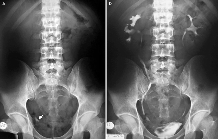
Fig. 4.14
Partially obstructing right distal ureter stone (arrow) on plain film (a) and as a filling defect on IVP (b)
Ureteral stones may cause urinary system obstruction, especially when they lodge into the physiologically narrower segments of the ureter (Fig. 4.15).
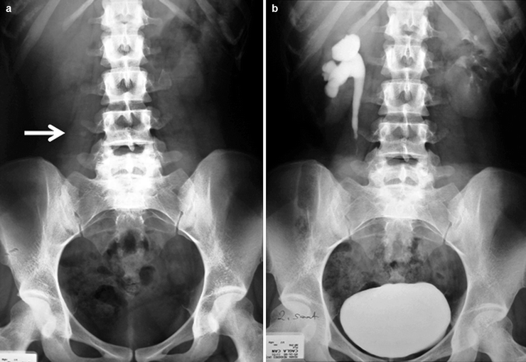
Fig. 4.15
Low density stone located in right ureter (a) arrow is accompanied by delay in renal function and hydronephrosis (b)
Obstruction leads to dilatation of the urinary tract above the calculus and delay in renal nephrogram (Fig. 4.16).
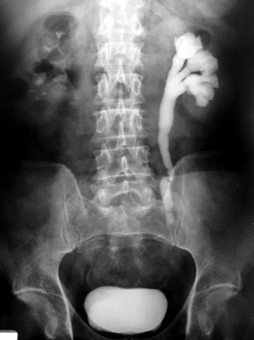
Fig. 4.16
Moderate hydroureteronephrosis and delayed renal function secondary to left ureter stone located at level of iliac vessel crossing
Ideally, the diagnostic quality of an IVP examination should be improved by bowel preparation to eliminate artifacts related to bowel contents because the potential hazards of contrast material injection, such as anaphylaxis and nephrotoxicity, and the use of ionizing radiation, weigh against repetition of the examination (Fig. 4.17).
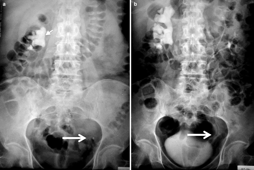
Fig. 4.17
KUB (a) and 25 min (b) IVP spot films of poor diagnostic quality. The relationship of the left side suspicious radiopacity in the bony pelvis and the bifid left ureter is visible (arrows). In addition, the right renal staghorn calculus (short arrow) is occluding the pelvicalyceal system
Abdominal compression maneuver can be used to improve the quality of IVP but should not be used in patients with abdominal aortic aneurysm.
Films taken in erect or prone positions may facilitate distal ureteral filling.
Obstructing stones cause delayed renal function; contrast passage distal to the stone is not readily seen, necessitating late spot films to attempt to evaluate renal function and to display the distal ureters.
Ultrasound
One of the widely used imaging modalities in the diagnosis of urinary calculus disease is ultrasound.
Urinary stones appear as echogenic foci with prominent posterior acoustic shadowing because the sound waves are strongly reflected by the surface of the stone (Fig. 4.18).
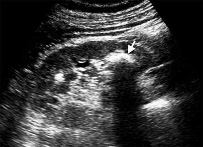
Fig. 4.18
Longitudinal gray-scale sonogram of the left kidney. Hyperechoic stone with prominent posterior acoustic shadowing (arrow) is present in the lower calyx
Millimetric calyceal stones or nonobstructing ureteral stones are difficult to detect with ultrasound.
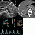
Doppler ultrasound may facilitate the diagnosis of small renal calculus if the “twinkle artifact” is present behind the suspicious echogenic focus (Fig. 4.19).
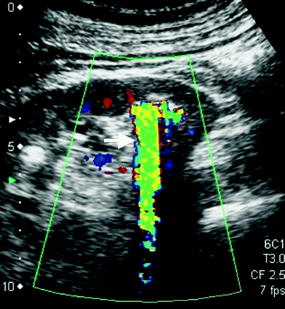
Fig. 4.19
Color flow Doppler ultrasound of the left kidney. Twinkle artifact (arrow) is present in the lower calyx stone
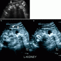
Dilatation of the pelvicalyceal system and ureters is an indirect sign of obstructing ureteric stone. Hydronephrosis can be graded by ultrasound (Figs. 4.20, 4.21, 4.22, and 4.23).
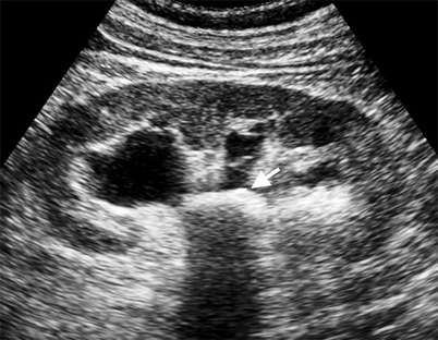
Fig. 4.20
Longitudinal sonogram of the kidney. Pelvic stone (arrow) is obstructing the collecting system; all calyces are moderately dilated
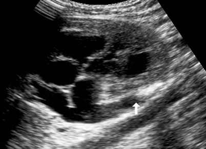
Fig. 4.21
Proximal ureter stone (arrow) is occluding the ureter, which in turn results in moderate hydronephrosis and clubbed calyces
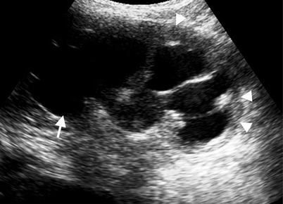
Fig. 4.22
Obstructing ureteric stone (not seen on this section) is causing severe hydronephrosis (arrow). Renal parenchymal thinning (arrowheads) secondary to hydronephrosis can be appreciated
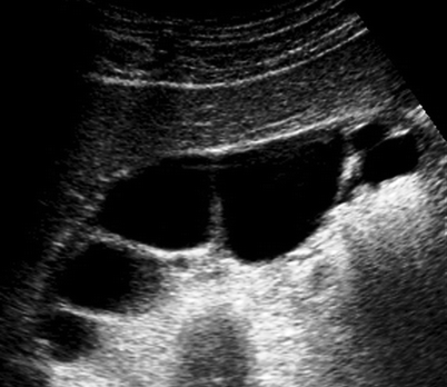
Fig. 4.23
Marked hydronephrosis of the kidney. Renal parenchyma is almost atrophied
Perinephric fluid collection may accompany urinary obstruction, secondary to forniceal rupture.Related posts:

Stay updated, free articles. Join our Telegram channel

Full access? Get Clinical Tree