KEY FACTS
Imaging
- •
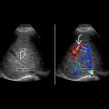
Dilated renal pelvis and calyces ± dilated ureter
- •
Distended bladder may cause functional obstruction or reflux resulting in hydronephrosis
- •
Low-level echoes within lumen suggest pus (pyonephrosis) or blood (hemonephrosis)
- •
Highly echogenic shadowing intraluminal structures represent stones, twinkling artifact on color Doppler
- •
Urothelial thickening suggests infection or rejection
- •
Ultrasound is sensitive and specific for hydronephrosis but may be limited for site of obstruction
Top Differential Diagnoses
- •
Nonobstructive dilatation, early postoperative edema
- •
Functional obstruction from overdistended bladder
- •
Prominent hilar vessels
- •
Renal sinus cysts (more common in native kidneys)
Pathology
- •
Causes include ischemic stricture, rejection, clot, calculus, extrinsic compression, infection, and tumor
- •
Reflux, infection, and decreased ureteral tone may cause nonobstructive dilatation
Clinical Issues
- •
Ureteral obstruction occurs in 3-6% of renal allografts
- •
Most common in first 6 months after transplantation
- •
Over 90% of strictures at ureterovesical anastomosis and distal 1/3 of ureter
Scanning Tips
- •
Look for transition zone and cause for obstruction
- •
Use color Doppler to distinguish hilar vessels from dilated renal pelvis and to distinguish clot or debris from solid tumor
- •
Use color Doppler to look for ureteral jet
- •
If bladder is distended, rescan with empty bladder
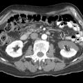
 in a renal transplant. The ureter
  is dilated secondary to obstruction by a fluid collection
  , which wrapped around the ureter.


