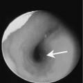
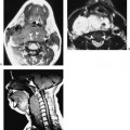
Chapter 133 Rouvière described a lateral and medial group of retropharyngeal lymph nodes (RPLNs) (Fig. 133–1). Primary squamous cell carcinoma arising from the nasopharynx, oropharynx, and hypopharynx may metastasize to the RPLNs. The most common primary malignancy that spreads to the RPLNs is nasopharyngeal carcinoma. More than 51% of patients with nasopharyngeal carcinoma will show RPLN involvement at presentation. The RPLNs are first echelon nodes of the nasopharynx, oropharynx, and hypopharynx. However, in one third of patients lymphatic drainage of the nasopharynx bypasses the first echelon nodes to drain directly into the internal jugular chain. Enlargement of the RPLNs is a radiological diagnosis because these nodes cannot be palpated. Patients with malignant nodes usually do not have symptoms referable to the metastatic nodes. Retropharyngeal lymph nodes should be considered as cervical lymph nodes when staging the neck. Hasegawa and Matsuura reported nodal size with histopathological proof in 24 patients with advanced carcinoma of the oropharynx and hypopharynx. The average size of 12 positive nodes was 15 mm (range 5–22 mm). The average size of 17 negative nodes was 11 mm (range 3–25 mm). The authors also reported that in 11 nodes > 15 mm, eight (73%) were positive, whereas in 18 nodes < 15 mm, only four (22%) were positive. There was clearly a large overlap of positive and negative nodes.
Retropharyngeal Lymphadenopathy
Epidemiology
Clinical Findings
Pathology
Treatment
Related posts:


Stay updated, free articles. Join our Telegram channel

Full access? Get Clinical Tree


