Clinical Presentation
The patient is a 76-year-old woman who presented with high cervical neck pain and headache. These symptoms have been ongoing for 6 months. There are no radicular features or myelopathic symptoms. There was no antecedent trauma. On physical examination, there is moderate limitation of neck movement in all directions. The patient is also noted to have moderate degenerative changes in both hands.
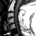
Imaging Presentation
Sagittal fat-saturated, T2- and T1-weighted images demonstrate soft tissue proliferation around the dens, more prominent posteriorly, with some mild mass effect upon the upper cervical spinal cord. There is some increased T2 signal intensity along the margin of the soft tissue. The findings are compatible with pannus formation with some surrounding bone marrow edema and/or inflammation ( Fig. 61-1 ) .

Discussion
Rheumatoid arthritis (RA) is a common systemic autoimmune process that causes chronic inflammation of the joint. It was first described by Garrod in 1890. A distinguishing feature of RA is symmetric and erosive synovitis of peripheral joints. RA has a predilection for cervical spine involvement and is the most common inflammatory disorder to affect the spine. RA never involves the spine without concomitant involvement of the hands and/or feet. It is estimated that RA affects approximately 1.3 million adults in the United States, with a worldwide prevalence of approximately 0.5% to 1% in developed countries. RA is more common in women by a ratio of 2 to 3:1, and although men are the minority of cases, they have a greater risk of more advanced cervical disease. Other risk factors that are associated with progressive disease include rheumatoid factor seropositivity, severe peripheral disease, and the prolonged use of steroids. RA demonstrates an increasing prevalence with older age, but interestingly, has been showing a progressive decrease in incidence in the younger age groups since the 1960s, making RA more and more a disease of older adults. Despite how common RA is, the etiology of this process remains unclear. Currently, it is thought that RA is an immune response to an antigenic expression by synovial cells.
Neck pain is a common symptom in patients with RA. The neck pain is often found at the craniocervical junction and may be associated with occipital headaches. The incidence of neck pain ranges from 40% to 88%. Other symptoms include mastoid pain, ear pain, migraine, and facial pain from compression of local nerves. There may also be myelopathic symptoms including weakness, gait disturbance, and paresthesia of the hands. If there is basilar invagination, the patient can present with vertigo, loss of equilibrium, or tinnitus.
The most serious complication of RA in the cervical spine is compression of the spinal cord or brainstem. This can occur from either subluxation of the spine or from direct pressure from pannus formation. Pannus formation around the dens may loosen or destroy the ligamentous structures, particularly the transverse ligament of the atlas and to a lesser extent the alar ligaments and apical dental ligament. This can lead to atlantoaxial subluxation, or horizontal instability, which is defined as a greater than 3 mm distance between the midpoint of the posterior aspect of the anterior arch of the atlas and the anterior aspect of the dens ( Fig. 61-2 ) . There can also be vertical instability, leading to basilar impression in which the dens extends through the foramen magnum ( Fig. 61-3 ) .


The upper cervical spine is most commonly affected in RA for two reasons: (1) the C0-C1 and C1-2 articulations are purely synovial and are therefore primary targets for RA, and (2) the C1-2 facet joint is oriented in the axial plane, and therefore, there is no bony interlocking to prevent subluxation. Cervical subluxations have been reported in 40% to 80% of patients on radiographs; however, neurologic deficits are seen in only 10% to 30% of patients.
The diagnosis of rheumatoid arthritis is made by a combination of clinical, physical, and/or radiologic findings as delineated in the revised 1987 Criteria for the Classification of Acute Arthritis of Rheumatoid Arthritis by the American Rheumatism Association. A patient must satisfy four of the seven criteria, and criteria 1 to 4 must have been present for at least 6 weeks.
- 1
Morning stiffness —Morning stiffness in and around the joints, lasting at least 1 hour before maximal improvement
- 2
Arthritis of three or more joint areas —At least three joint areas simultaneously have had soft tissue swelling or fluid (not bony overgrowth alone) observed by a physician. The 14 possible areas are the right or left proximal interphalangeal joint, metacarpal phalangeal joint, wrist, elbow, knee, ankle, and metatarsal phalangeal joints
- 3
Arthritis of hand joints —At least one hand joint area swollen (as defined in #2)
- 4
Symmetric arthritis —Symmetric to nearly symmetric simultaneous involvement of the same joint areas (as defined in #2) on both sides of the body
- 5
Rheumatoid nodules —Subcutaneous nodules over bony prominences, extensor surfaces, or in juxtaarticular regions
- 6
Serum rheumatoid factor —Demonstration of abnormal amounts of serum rheumatoid factor by any method
- 7
Radiographic changes —Erosions or unequivocal bony decalcification of the hands or wrists
Related posts:


Stay updated, free articles. Join our Telegram channel

Full access? Get Clinical Tree


