Ruptured AVM
Christopher J. Karakasis
CLINICAL HISTORY
11-year-old female presenting with the worst headache of her life, which began abruptly after jumping up and cheering at a basketball game. She quickly developed left-sided weakness and numbness.
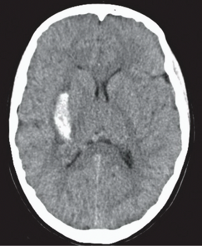 FIGURE 77A |
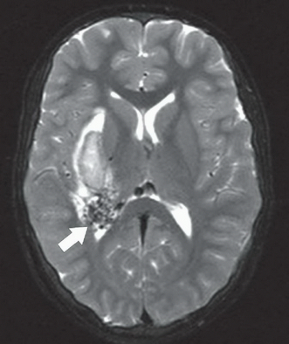 FIGURE 77B |
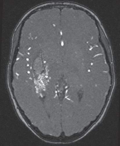 FIGURE 77C |
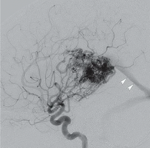 FIGURE 77D |
FINDINGS
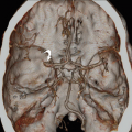
Figure 77A: Noncontrast head CT demonstrates acute hemorrhage centered in the posterolateral putamen and external capsule. Note also the subtle area of subtle hyperdensity just posterior to the hemorrhage. Figure 77B: Axial T2WI demonstrates a cluster of hypointense flow voids in this area (arrow) posterior to the hematoma. Figure 77C: Axial source image from a 3D-TOF MRA demonstrates punctate and serpiginous flow-related enhancement in the area posterior to the hematoma, suggesting arterial flow. Figure 77D: Lateral projection from cerebral angiography with injection of the right internal carotid artery demonstrates a tangle of abnormal vessels supplied by perforators from the right middle cerebral artery and anterior choroidal artery, as well as early filling of the straight sinus (arrowheads).
Related posts:


Stay updated, free articles. Join our Telegram channel

Full access? Get Clinical Tree


