Fig. 23.1
Sacrectomies performed above the S1 neural foramen (a) were found to be unable to withstand postoperative mobilization compared to osteotomies performed below the S1 level (b). Due to these findings, reconstruction is recommended when an osteotomy is performed cranial to the S1 foramen
23.3 Spinopelvic Reconstruction
In order to restore spinal balance, and to reconstruct the spinopelvic instability that is generated by the sacrectomy, pedicle and iliac screw fixation with posterior rods and anterior support is currently our standard technique. Historically, the use of spinal instrumentation was associated with a high failure rate related to screw loosening and rod breakage, likely due to the use of posterior-only spinal reconstruction techniques without anterior column support at the resected sacral level. However, newer combined anterior and posterior reconstruction techniques have reduced failure rates [9, 10].
The ilium has two pathways of abundant cancellous bone: the upper (posterior superior iliac spine to the iliac crest) and lower segments (posterior superior spine to the anterior inferior iliac spine) which are ideal sites for anchoring iliac screws. It has been shown that if iliac screws are placed in both these paths, there is significant resistance to both compressive and torsional forces, especially with dual lower segment fixation [11]. In addition to improved iliac fixation techniques, changes have occurred in posterior rod instrumentation techniques, namely, the four-rod reconstruction technique. In an in vitro total sacrectomy model, Kelly and colleagues were able to demonstrate the superiority of a four-rod reconstruction technique compared to the traditional two-rod construct to provide significantly greater stability in flexion and extension [12]. Likewise, the addition of cross-links to the four-bar construct significantly increased the rotational stability of the construct [12]. In addition to a four-rod reconstruction with cross-linking, our preferred reconstruction technique includes an anterior column reconstruction of the sacral vertebral segment to augment the posterior reconstruction with anterior strut grafts (which are often preferably vascularized free fibulas), forming a “cathedral-shaped” reconstruction of the anterior spinal column at the sacral level [8].
23.3.1 Indications for Reconstruction
Reconstruction at our institution is performed when the SI joint has been disrupted, based on the Mayo Spino-Sacral-Pelvic Tumor Classification (Fig. 23.2), following either a total sacrectomy (Mayo Type IA), partial sacrectomy above the S1 foramen (Mayo Type IB), unilateral hemisacrectomy (Mayo Type II), amputative partial sacrectomy plus hemipelvectomy (Mayo Type III), or total sacrectomy and hemipelvectomy (Mayo Type IV). Reconstruction is not necessary for a lower sacrectomy which does not disrupt the SI joints (Mayo Type IC).
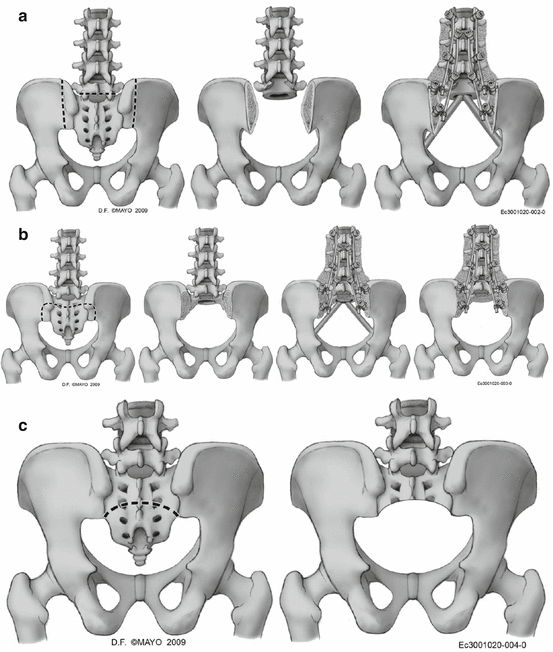
Fig. 23.2
The Mayo Spino-Sacral-Pelvic Tumor Classification assists in the planning of reconstruction based on the extent of resection and also if there is an amputative pelvic procedure. We perform a spinopelvic reconstruction for cases of Mayo Type IA (total sacrectomy, a), Type IB (subtotal sacrectomy above the S1 foramen, b), Type II (hemisacrectomy involving the ipsilateral sacroiliac joint, d), Type III (hemipelvectomy and hemisacrectomy, e), and Type IV (total sacrectomy and hemipelvectomy, f). Since the SI joints are not disrupted in a Type IC (subtotal sacrectomy below the S1 foramen, c), a reconstruction is not typically performed
23.3.2 Cathedral Reconstruction
Staging of a total sacrectomy (Mayo Type 1A) has been shown to reduce patient morbidity and is currently our preferred technique [13]. We perform this procedure in three stages on three different surgical days. The anterior procedure, which we perform on day 1, includes the harvesting of a vertical rectus abdominis vascularized myocutaneous (VRAM) flap; the pelvic viscera mobilization; the sacral, spinal, and pelvic osteotomies as needed for a specific patient; the vascular mobilization; the nerve root sectioning as needed for a specific patient’s tumor geometry; and the positioning of the VRAM flap in the pelvis over a Silastic sheet that identifies the plane of pelvic dissection. The second stage is the posterior approach and dissection, completion of osteotomies, tumor delivery and removal from the surgical field, and provisional VRAM flap inset followed by wound closure. The patient gets a spinopelvic CT scan on the way to the intensive care unit, which is used in the planning for the reconstruction which occurs as stage 3. The stage 3 reconstruction consists of bilateral bicortical pedicle screws in the two or three most distal remaining vertebrae and one or two iliac screws on each side. Docking sites for the two fibulae which constitute the anterior column sacral reconstruction are then created with a burr in the most caudal portion of the lowest remaining vertebra and in the ilia. The iliac docking sites may be prepared in the supra-acetabular region, in the ischial ramus, or in the ischial tuberosity area, depending on the locations that remain after specimen removal, as seen on the post-resection CT scan. The pelvic docking sites may be prepared during the anterior stage 1 operation, the posterior stage 2 tumor removal operation, or the stage 3 reconstruction operation, depending on the particular patient’s tumor anatomy and which of the three operations present the optimum opportunity to prepare the docking sites. The ideal position of the docking sites in the ilium is at the intersection of the iliopectineal line and a straight line which intersects the center of the hip joint and the most caudal remaining vertebra (Fig. 23.3a). The position of these docking sites ultimately depends on the location and extent of the tumor removal in each patient and the bone areas that remain and are available for fibular strut graft docking.

Fig. 23.3
During the anterior exposure of the tumor, receptacles for the strut grafts are made on the iliopectineal line at the crossing point from the center of the planned distal vertebrae and the center of the hip joint. Following tumor extirpation, bicortical pedicle screws are placed in the remaining lumbar vertebrae, and a docking site is made in the central portion of the most distal vertebrae. The fibular strut grafts are then placed into the docking sites of the ilium (a) and then inserted into the distal vertebrae in a “cathedral” fashion (b). Compression is then placed across the docking site, and the nuts of the pedicle screws are tightened (c)
Fibular allografts or autografts are fashioned in order to be inserted into the docking sites, creating a “cathedral” appearance (Fig. 23.3b). We currently advocate the use of vascularized free fibulas instead of allografts in the setting of previous sacropelvic radiation. In such patients, healing of an allograft may likely be compromised. Following the docking of the fibulae to the most distal remaining vertebra, a bilateral four-rod posterior instrumentation reconstruction with cross-links is performed, and the instrumentation construct is compressed across the docking site (Fig. 23.3c).
Related posts:


Stay updated, free articles. Join our Telegram channel

Full access? Get Clinical Tree


