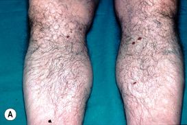
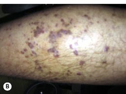
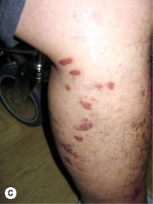
Figure 20.28A–C). These tumours develop slowly, but patients are at risk of developing a second malignancy, often non-Hodgkin’s lymphoma. Most patients are treated with relatively low doses of radiotherapy, such as 8 Gy in a single fraction, which may be repeated.
Stay updated, free articles. Join our Telegram channel

Full access? Get Clinical Tree


