Presentation and Presenting Images
( ▶ Fig. 9.1, ▶ Fig. 9.2, ▶ Fig. 9.3, ▶ Fig. 9.4)
A 42-year-old female whose mother had postmenopausal breast cancer presents for screening mammography.
9.2 Key Images
( ▶ Fig. 9.5, ▶ Fig. 9.6, ▶ Fig. 9.7, ▶ Fig. 9.8, ▶ Fig. 9.9, ▶ Fig. 9.10, ▶ Fig. 9.11)
9.2.1 Breast Tissue Density
The breasts are heterogeneously dense, which may obscure small masses.
9.2.2 Imaging Findings
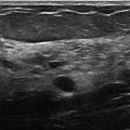
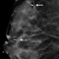
Scattered throughout both breasts, there are multiple masses of variable size. On the digital breast tomosynthesis (DBT) images at least two-thirds of the margins of the masses are circumscribed and the remaining margins are obscured ( ▶ Fig. 9.5, ▶ Fig. 9.6, ▶ Fig. 9.7, and ▶ Fig. 9.8). The patient had an ultrasound last year ( ▶ Fig. 9.9, ▶ Fig. 9.10, and ▶ Fig. 9.11) that demonstrated multiple simple cysts (anechoic masses with circumscribed margins and posterior acoustic enhancement).
9.3 BI-RADS Classification and Action
Category 2: Benign
9.4 Differential Diagnosis
Multiple bilateral masses, benign: All of the masses have similar features with at least 75% of the margins circumscribed and the remaining margins obscured by adjacent fibroglandular tissue.
Fibroadenomas: Patients can have multiple bilateral masses that are fibroadenomas. These would be benign as long as they fit the criteria of bilateral benign-appearing masses. In this case, the prior ultrasound of this patient revealed that these masses are cysts.
Metastases: Metastases can occur to the breast and occasionally can be the presenting sign of an unknown cancer. However, this is much less common than a benign etiology. This patient has no history of a confirmed cancer.
9.5 Essential Facts
The BI-RADS classification of masses used for mammography should be similarly applied to DBT.
Multiple bilateral masses is defined as having at least three masses, with at least one in each breast. The masses are at least partially circumscribed, with at least 75% of the margins circumscribed and the remaining margins obscured by adjacent fibroglandular tissue.
9.6 Management and Digital Breast Tomosynthesis Principles
It is possible that DBT could greatly increase the cases that can be defined as multiple bilateral masses, because of DBTs ability to unmask the obscuring tissues and better define the underlying circumscribed masses.
Thus, DBT may increase the number of masses that can be assigned a BI-RADS 2 assessment.
A limitation of DBT can also be associated with an advantage of DBT. DBT decreases the effect of overlapping tissue, thus benign lesions that were previously concealed will be detected. Additional further evaluation may be prompted by this unmasking.
9.7 Further Reading
[1] Leung JW, Sickles EA. Multiple bilateral masses detected on screening mammography: assessment of need for recall imaging. AJR Am J Roentgenol. 2000; 175(1): 23‐29 PubMed
[2] Roth RG, Maidment AD, Weinstein SP, Roth SO, Conant EF. Digital breast tomosynthesis: lessons learned from early clinical implementation. Radiographics. 2014; 34(4): E89‐E102 PubMed

Fig. 9.1 Right craniocaudal (RCC) mammogram.
Stay updated, free articles. Join our Telegram channel

Full access? Get Clinical Tree


