Chapter 173
Schwannoma
Epidemiology
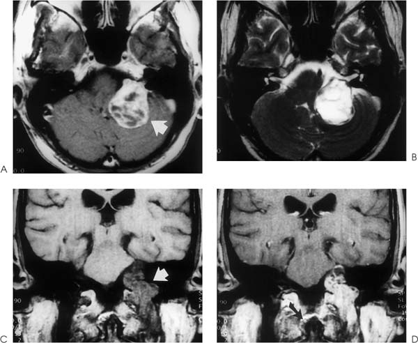
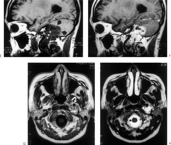
Schwannomas most commonly affect cranial nerve VIII. In the neck, they usually involve the vagus nerve but may also originate in other cranial nerves and the sympathetic trunk. Schwannomas are more common neurofibromas. They usually present in the fourth decade of life.
Clinical Presentation
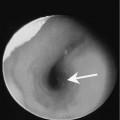
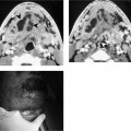
Schwannomas in the neck may present as a painless mass. Deeply seated tumors in the region of the jugular foramen may cause nerve compression resulting in single or multiple cranial nerve palsies.
Pathology
Schwannomas, unlike neurofibromas, are encapsulated tumors. Histology shows Schwann’s cells organized in compact interlacing groups associated with fibrous strands. Schwannomas may show two histological patterns. Antoni type A shows compact palisading cells whereas Antoni type B shows a less cellular pattern consisting of cells with cytoplasmic lipids. Cystic degeneration is common. These tumors arise from a single focus and they grow by projecting from one side of the nerve.