Abstract
Scimitar syndrome is a rare congenital pulmonary anomaly characterized by a unilateral anomalous pulmonary venous drainage consisting of an aberrant vein that drains the lung blood flow into the systemic venous circulation (usually the inferior vena cava) and is associated with an ipsilateral pulmonary hypoplasia, underdevelopment of the ipsilateral pulmonary artery, and sometimes an anomalous systemic arterial supply from the descending aorta to the hypoplastic lung. The diagnosis can be established during fetal life by ultrasound identification of anomalous drainage of any pulmonary vein together with ipsilateral lung hypoplasia and mediastinal shift. It is usually a nonlethal anomaly, but the prognosis depends on the degree of lung hypoplasia and the presence of a systemic feeding artery to the hypoplastic lung, which can lead to neonatal pulmonary hypertension with a left-to-right cardiac shunt.
Keywords
scimitar syndrome, pulmonary venolobar syndrome, hypogenetic lung syndrome
Introduction
Scimitar syndrome (SS) is a rare congenital anomaly characterized by a partial (usually the right lower lobe) or complete unilateral anomalous pulmonary venous drainage in association with ipsilateral lung hypoplasia. The anomaly, also known as pulmonary venolobar syndrome or hypogenetic lung syndrome , was first described in 1836. The term scimitar was first used by Halasz et al. to describe the radiographic appearance of the anomalous pulmonary vein trajectory that drains into the inferior vena cava (IVC) just below or above the right hemidiaphragm as a broad, gently curved shadow resembling the silhouette of a short, curved Persian sword known as a shimshir . In 1960, the familial occurrence and the clinical spectrum were defined first using the term scimitar syndrome .
Disorder
Definition
SS is a congenital pulmonary anomaly consisting of an anomalous vein that drains part or all of the right lung blood flow into the systemic venous circulation, usually the IVC, associated with an ipsilateral pulmonary hypoplasia, underdevelopment of the right pulmonary artery, and sometimes an anomalous systemic arterial supply from the descending aorta to the hypoplastic lung. Although SS has been classically described as involving the right lung, this might be due to the extreme rarity of a left-sided vena cava, because some case reports of left-sided SS have also been described.
Prevalence and Epidemiology
Most cases reported were in adults or older children, accounting for 1 : 2000 cases of congenital heart disease. Only a few prenatal cases have been reported, with an estimated incidence of 1 : 100,000 to 3 : 100,000 live births and a clear female predominance.
Etiology and Pathophysiology
Isolated SS is usually sporadic and not associated with chromosomal or genetic abnormalities, but in the presence of associated malformations, the risk of chromosomal defects increases to 50%. Association with other malformations is common, including congenital heart disease in 75%, lung congenital cystic malformations, congenital diaphragmatic hernia (CDH), and vertebral anomalies. The most commonly reported congenital heart malformation is persistent left superior vena cava draining to the coronary sinus ; other abnormalities that have been described include tetralogy of Fallot, coarctation of the aorta, abnormalities of the aortic arch, atrial septal defects, absent IVC, and pulmonary vein stenosis contralateral to the hypoplastic lung.
The pathogenetic mechanism of SS is unclear. It seems to originate from an abnormal lung development during embryogenesis. It has been hypothesized that connections between the pulmonary and systemic plexus of capillaries normally present during embryonic development remain patent, leading to pulmonary hypoplasia and underdevelopment of the ipsilateral pulmonary artery. There is no evidence of the influence of common teratogens in the genesis of SS.
Manifestations of Disease
Clinical Presentation
There are often no prenatal manifestations. Hypoplasia of the right lung may cause a rightward shift of the heart. SS is usually a nonlethal anomaly. Although extremely rare, coexistence with stenosis of the pulmonary vein contralateral to the hypoplastic lung may be associated with a high neonatal mortality rate. In the absence of additional congenital malformations, the prognosis depends on the degree of lung hypoplasia and the presence of abnormal arterial blood supply to the affected lung, which can lead to pulmonary hypertension with a left-to-right cardiac shunt in 50% of the cases. In cases with mild pulmonary hypoplasia and an absence of any abnormal systemic artery supplying the right lung, the prognosis is normally good. In many instances, the condition is asymptomatic after birth and remains clinically silent for a long time, sometimes to adulthood. In more severe cases manifesting in infancy and childhood, common clinical manifestations are dyspnea, cyanosis, respiratory distress or cardiac failure owing to increased pulmonary blood flow secondary to the aberrant pulmonary arterial supply, and recurrent respiratory tract infections during infancy.
Diagnosis
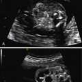
Prenatal ultrasound (US) is characterized by anomalous drainage of any pulmonary vein ( Figs. 5.1 and 5.2 ) together with ipsilateral lung hypoplasia and mediastinal shift. SS can usually be detected on US during the second trimester of pregnancy. The anomalous venous drainage is usually into the IVC below the diaphragm but may join with the hepatic vein, portal vein, coronary sinus, right atrium, or more unusually, with the superior vena cava or the left atrium.


Stay updated, free articles. Join our Telegram channel

Full access? Get Clinical Tree


