Syndromes
Vascular malformations
Venous
Capillary
Lymphatic
Arterial or arteriovenous
PHACES
LUMBAR
•
Kasabach–Merritt
•
Maffucci
•
•
Klippel–Trenaunay
•
•
•
•
Gorham–Stout
•
•
•
Proteus
•
•
•
Parkers Weber
•
•
•
Sturge–Weber
•
•
CM-AVM
•
•
Familial cerebral VM
•
Familial cutaneomucosal VM
•
Blue rubber bleb nevus
•
Bockenheimer
•
Cobb
•
•
Osler–Weber–Rendu
•
•
Beckwith–Wiedemann
•
Louis–Barr
•
Down
•
Turner
•
Wyburn–Mason
•
Dandy–Walker
Von Hippel–Lindau
Natural History/Epidemiology
VMs are present at birth. They are clinically not always apparent and tend to grow in proportion to the growth of the child. The growth is most pronounced during puberty and pregnancy. These are congenital lesions that affect boys and girls of equal frequency with a reported incidence of 1–2 per 100,000 births and a prevalence of 1 % [7].
Most VMs (95 %) are sporadic, but can be seen in a number of heritable conditions. The molecular basis for sporadic is yet to be discovered, however there are familial cases where the genetic defect has been localized on a specific chromosome. In 1994–1995, two families were identified to have autosomal dominant inherited cutaneous and mucosal VMs. Genetic analysis mapped a locus for both of these families to the chromosome 9p21 [8, 9]. These families shared a mutation resulting in an arginine to tryptophan substitution R849W in the gene that encodes the kinase domain of the endothelial cell receptor Tie2 [10]. In 1999, four more families with autosomal dominant inherited VMs were identified [11]. Only one of those families shared the same mutation as the previous two reports. The second family had a novel hyperphosphorylating Y897S mutation in the TIE2 gene. The other families showed no evidence of linkage to 9p21, which suggests genetic heterogeneity. Multifocal VMs are most commonly seen in the familial forms of VM, including the following: Blue rubber bleb nevus (BRBN) syndrome, Mucocutaneous familial VMs, Glomovenous malformation, and Maffucci’s syndrome [12].
Diagnostic Imaging
Ultrasound: On gray-scale imaging, VMs usually are hypoechoic to anechoic with the shape of tubular structures [13]. Some lesions can have a heterogeneous echotexture if phleboliths or different forms of thrombus are present within the lesion. Doppler ultrasound is usually a monophasic low-velocity waveform. Sometimes flow can only be seen with compression and release of the lesion [14].
Computed tomography: On non-contrast CT, VMs are usually hypoattentuating, however, they can be heterogeneous depending on the amount of fatty tissue within the lesion. Phleboliths or dystrophic calcifications can be seen within the lesion. After the administration of contrast, the lesion usually enhances at the periphery and then fills in centrally on the delay images [14]. CT is excellent at looking for boney involvement from the lesion. MRI is better at characterizing the lesions relationship to the surrounding soft tissue structures .
MRI: VMs appear usually as hypointense to isointense on T1-weighted imaging. They can however have areas of bright signal on T1-weighted images if the lesion has fat, subacute blood products, or certain types of calcifications contained within the lesion [15]. On T2-weighted images, VMs have high signal intensity. Gradient echo sequence usually shows areas of low signal corresponding to calcification, hemosiderin, or thrombus. The best sequence to determine the fully extent of the lesion and its relationship to surrounding soft tissue structures is the T2-weighted sequence [16]. On T1-weighted post contrast imaging, the lesions usually demonstrates early peripheral enhancement that later fills in centrally on delayed imaging [17].
Diagnostic venography: Contrast venography is helpful in the anatomical characterization of a VM. It is performed to determine the fully extent of the malformation and its draining veins. It is also helpful in confirming patency of the deep venous system in the extremity. Venography is also performed to determine the volume of contrast needed to fill the malformation before empties into normal draining veins .
Based on the pattern of venous drainage Dubois and Puig divided VMs into four types [18–20]. This drainage pattern is a reflection of the response to the treatment and rates of complications. Types I and II respond best to sclerotherapy with a better control rate and a lesser number of sessions to achieve control. Types III and IV have a higher rate of complications [20] .
Type I: isolated malformation without disconcernible venous drainage
Type II: lesion draining into normal veins
Type III: lesion draining into dysplastic veins
Type IV: lesion consists primary of venous ectasia
Treatment
VMs can be treated percutaneously by injecting a sclerosing agent. The most commonly described sclerosing agents (Table 22.2) included: alcohol, sodium tetradecol, ethibloc, polidocanol, and bleomycin [19, 21–24]. The procedure is performed under general anesthesia, since injection of the sclerosing agent can be extremely painful. A foley catheter is placed in the bladder to monitor urine output and color, since some patients may develop a hemoglobinuria. This is most likely to happen in younger patients and in patients with large lesions. In addition, if an extremity is being treated, we start an IV distal to the lesion to infusion saline and heparin overnight to flush out the sclerosing agent and prevent acute deep venous thrombosis [12]. Sclerotherapy is performed by cannulating the lesion percutaneously under ultrasound or fluoroscopy. Contrast material is injected to define the extent of the lesion, determine the flow rate within the lesion and the communication with normal veins, the presence of a possible arterial component, and to give an estimate of the volume of sclerosing agent needed to fill the lesion. If a large draining vein is visualized, these can be treated with embolization utilizing coils, liquid embolic agents, or with temporary balloon occlusion to increase the contact time of the sclerosing agent with the VM and to prevent egress of the agent into the systemic circulation [12]. This may also be performed with a tourniquet, blood pressure cuff, or manual compression. The VM is accessed with ultrasound guidance and the sclerosant is then injected under fluoroscopic guidance to decrease the risk of extravasation, overfilling of the lesion, and limit undesired egress of the sclerosing agent into the normal deep veins (Fig. 22.1). Overfilling of the lesion may also be prevented by the two-needle access technique, which allows decompression of the lesion through the second needle access. Cone beam CT can be performed to evaluate the anatomical distribution of the administered sclerosant and compare the distribution to the preprocedure imaging [25]. After the needles are removed, a 20–30 mmHg compression stocking is placed over the area of concern so that the veins do not refill with blood. This allows maximum contact of the damage circumference endothelium of the vein walls, promoting fibrosis and scarring. The procedure is then repeated at 8–10 weeks intervals, until there is no recanalization, swelling, or pain from the lesion. Diffuse VMs are much more difficult to treat. The area of maximum symptomatology is usually targeted, since these lesions cannot be completely obliterated .
Table 22.2
Common sclerosing agents
Alcohol |
Sodium tetradecol |
Bleomycin |
Doxycycline |
Alcoholic solution of zein (Ethibloc) |
Polidocanol |
OK-432 (Picibanil) |
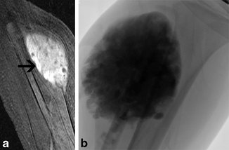
Fig. 22.1
A 4-year-old boy who complains of pain and swelling within his left calf from a venous malformation. a Coronal post contrast fat saturation T1-weighted MRI image shows an enhancing mass within the left calf (black arrow) with a few scattered areas of round low signal within it (small black arrow), consistent with pheboliths. b Frontal spot fluoroscopic image shows the venous malformation filled with alcohol mixed with contrast
After sclerotherapy, the lesion should feel firm to palpation. Swelling is generally maximum 24 h after the procedure. We recommend placing a custom fitted class II compression garment over the lesion as soon as possible. If the lesion involves the airway, the patient may need to be kept intubated overnight in the ICU. The patient is given IV steroids in the hospital and then sent home on a tapered dose pack. The affected area is kept elevated above the heart with ice packs applied to minimize swelling. Patients receive IV fluids twice the maintenance dose before, during, and after the sclerotherapy for 24 h. Urine output is closely monitored for hemoglobinuria. Appropriate IV and oral analgesics and anti-inflammatory agents are prescribed in the hospital and post discharge for 7 days afterwards.
Patients with extensive limb lesions should be instructed from childhood in the proper use of compression garments. Compression helps to decrease the discomfort associated with the lesion, protects the overlying skin, limits swelling, and improves localized intravascular coagulation.
In patients with extensive VMs in whom a low fibrinogen is present, the use of low molecular weight heparin is recommended for 2 weeks pretreatment and possible cryoprecipitate transfusion if still low on the day of the procedure. This therapy will reduce the consumptive coagulopathy of the extensive VM and potentially lower the recanalization rate [12] .
Sclerosant Drugs
Absolute ethanol (95–98 %) is probably the most common agent used for sclerotherapy. It is the most effective sclerosant agent available, however it is also the most toxic [12]. Ethanol works by causing instant precipitation of endothelial cell proteins and rapid thrombosis (Fig. 22.2). It may also cause transmural vessel necrosis resulting in diffusion into the surrounding tissues. Serious side effects from ethanol injection include: massive swelling, tissue necrosis, hypoglycemia, peripheral nerve injury, CNS depression, hemolysis, pulmonary vasospasm, cardiac arrhythmias, and electromechanical disassociation [26–33]. Given the serious side effects, patients must be closely monitored during ethanol procedures. Some practitioners even suggest monitoring of pulmonary artery pressure during and for a short while after the procedure. Ethanol blood levels correlate directly with the amount of ethanol injected [34]. Mason et al. demonstrated that a level of > 1 ml/kg may put patients at increased risk of respiratory depression, cardiac arrhythmias , seizures and rhabdomyolysis [35]. A total dose of 1 ml/kg (or 60 ml) per session should never be exceeded. In pediatric patients, a maximum dose of 0.5 mg/kg per single session is recommended. Ethanol can be injected in an undiluted form, which we feel is dangerous since the agent cannot be identified under fluoroscopy. We usually dilute alcohol with water-soluble liquid or oily contrast medium during injection. Alcohol should not be mixed with Visipaque since this will cause precipitation of the contrast medium. In a series of 60 patients with VM involving the head and neck treated with ethanol, Su et al. reported a 68 % complete response (> 90 % volume reduction), 25 % marked response (> 50 % volume reduction), and a 7 % moderate response (< 50 % volume decrease) with a 10 % complication rate [36]. In a series of 158 patients with VM, 16 % of patients had a good response defined by a clinical examination and a ≥ 30 % decrease in the size of MRI, unfortunately 27 % of these patients experienced a complication [37]. In another series of 87 patients with craniofacial VMs treated with ethanol sclerotherapy, 32 % had an excellent response (> 75 % volume reduction), 52 % a good response (> 25 % volume reduction), and a 16 % a poor response (25 % volume decrease) with a 5 % complication rate [38] .
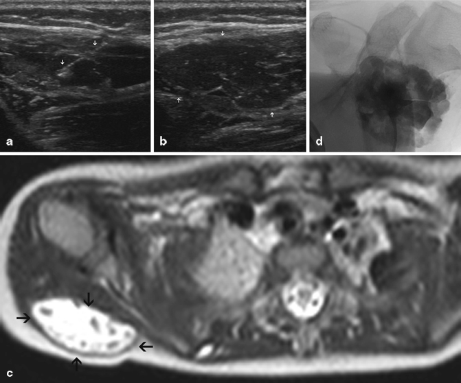
Fig. 22.2
A 3-year-old girl who complains of pain and swelling along with a bluish discoloration within the posterior right shoulder. a Gray-scale ultrasound image obtained with a liner 10 MHz transducer of the posterior right shoulder demonstrates a hypoechoic mass just below the cutaneous soft tissue (white arrows). b Ultrasound image shows an angiocatheter entering the mass (white arrows). c Axial T2-weight MRI image of the shoulder demonstrates a high-signal intensity mass with a few areas of low signal within it involving the soft tissue of the posterior right shoulder (black arrow). d Spot fluoroscopic image shows the venous malformation filled with alcohol mixed with contrast
Detergents
Detergents used for sclerotherapy of VM include sodium tetradecyl sulfate (STS), polidocanol, sodium morrhuate, and ethanolamine [39–43]. STS is approved in the USA for the treatment of varicose veins [44]. Polidocanol has an FDA approval for sclerosis of spider and reticular veins [45]. Similar to ethanol, all of the drugs damage the endothelial cells, resulting in thrombosis and fibrosis. Detergents may be mixed with water soluble or oily contrast medium prior to injection. These agents are thought to have a low rate of complications when compared to ethanol, but a greater tendency towards recanalization. Reported complications include cardiovascular collapse, skin pigmentation, skin necrosis, anaphylaxis, and hemoglobinuria [46]. Foaming the sclerosant by mixing the drug with air has also become popular [42, 47]. The theory is that the foam probably results in better contact between the drug and vein wall along with prolonged displacement of the blood with the lesion. Air can be made easily by mixing 10 cc of sclerosant with 3 cc of Ethiodol and 5–10 cc of air through a three-way stopcock. Patient receiving large volumes of detergent sclerosants may need aggressive hydration and alkalization of the urine to treat hemoglobinuria [39]. In prospective randomized controlled trial comparing foamed versus liquid polidocanol or ethanolamine in 89 patients with VMs, 90 % of patients had partial or complete response in the foamed group versus 63 % in the liquid group (p = 0.002) [48]. In another series of 50 patients with VMs treated with polidocanol foam resulted in complete resolution in 38 % of patients, size reduction of ≥ 50 % in 30 % of patients, and a size reduction of 0–50 % in 26 % of patients, and no change in 8 % of the patients [49]. Reported complications were noted in 8 % of the patients. Furthermore, in a series involving 26 patients with VM using ethanolamine oleate showed 49 % excellent, 39 % good, 12 % fair, and 0 % poor results based on the clinical examination [50].
Bleomycin
Bleomycin is an antibiotic and antitumor agent originally derived from Streptomyces verticillus in 1966, which causes an inflammatory response in endothelial cells [21]. Bleomycin can be mixed with water-soluble liquid or oily contrast medium. There is some concern with the use of this agent due to the association with pulmonary fibrosis in the setting of chemotherapy. The threshold dose for pulmonary fibrosis and interstitial fibrosis is 450 units, whereas doses for sclerotherapy are typically 0.5–1 units/kg for pediatric patients and 1–15 units in adults [21]. Reported complications include skin ulceration, skin pigmentation, laryngeal edema, flu-like symptoms, cellulitis, nausea and vomiting, and focal alopecia [21, 51]. In a series of 31 patients with VM treated with percutaneous injection of bleomycin resulted in 0 % complete resolution, 34 % marked (≥ 50 %) decrease in size, 31 % minimal (≤ 50 %) decrease in size, 34 % stable size, and 0 % increase in size on post procedure MRI [51]. Complications were seen in 12.5 % of patients comprised predominantly of transient skin pigmentation and cellulitis. Muir in another series of 32 patients treated with percutaneous bleomycin sclerotherapy showed complete resolution of the lesion in 32 % of patients and significant improvement in 52 % of patient by clinical examination [21].
Liquid Embolic Agents
N-butyl-2-cyanoacrylate (n-BCA) has a limited role in the treatment of vascular malformations because of its high cost, limited reabsorption, and formation of a hard mass after injection that may last for many months. Burrows et al. suggest this form of treatment for preoperative embolization prior to surgical resection [12]. This is most common in the treatment of intra-articular VMs where injection of a sclerosing agent could cause possible damage to the articular cartilage. This form of treatment has also been described for an orbital VM [52]. Ethylene vinyl alcohol copolymer (Onyx), a newer liquid polymer may also be useful for preoperative embolization of VMs prior to resection.
Other Forms of Treatment
Endovascular diode laser therapy has been reported in a small series of VMs with a good response rate at 14-month follow-up [53]. In patients with Klippel–Trenaunay syndrome, endovenous laser ablation of the marginal vein may be a useful alternative therapy. Percutaneous, interstitial (nonendovascular) laser photocoagulation of VM using the diode or Nd:YAG lasers via a fiber optic delivery system has also been reported with reasonable success [54–58]. Because of the controlled delivery of energy, and with external cooling, this modality may be of use in extensive superficial cutaneous/subcutaneous lesions or isolated anatomic locations such as the digit or tongue where sclerosis or embolization may carry a higher risk of surrounding tissue necrosis.
Surgical resection of VM is indicated only when complete resection without a resulting functional or anatomical deficit is possible. Surgery plays a limited role in the management of extremity lesion, but if performed localized coagulopathy must be controlled before surgery [19, 59, 60]. Surgery may also help in avoiding joint distention from repeat hemarthrosis, if joint involvement with the VM of the synovium is present.
Lymphatic Malformation
Clinical Features
LMs are present at birth and are composed of abnormal dilated lakes of lymphatic tissue that result from a defective embryological development of the primordial lymphatic channels. LMs can be classified radiographically as macrocystic (cysts ≥ 2 cm), microcystic (cysts < 2 cm), or mixed, which has important implications for treatment. Macrocystic lesions are most commonly located in the neck, axilla, and chest wall and when large may interfere with the birth process. Microcystic lesions usually present as diffuse soft tissue thickening, often associated with vesicles of the skin and mucosa with an overlying capillary malformation (CM) . The lesions grow in proportion to the patient. They may undergo periodic swelling often associated with signs of inflammation, which may be spontaneous or related to a regional infection. Acute swelling of the lesion may also be related to hemorrhage or lymphatic obstruction. Symptoms due to LMs are typically related to the localized mass effect on adjacent structures. On physical examination, LMs are mobile and have a boggy or cystic consistency. The lesions cannot be decompressed and do not distend with the Valsalva maneuver like VMs. The presence of lymphatic endothelium can be confirmed by staining with D2-40 antibody [61] .
Infection is usually common in suprahyoid LMs with mucosal involvement. Patient with airway involvement can present with stridor and sleep apnea. In one series, tracheostomy placement occurred in 5 % of patients with airway compromise at birth. Orbit LM can present with pain, swelling, proptosis, blepharoptosis, and emblyopia. An underlying cerebral developmental anomaly is seen in 45 % of patients with orbital involvement.
Natural History/Epidemiology
LMs are present at birth. Developmental defects during embryonic lymphangiogenesis result in LMs [7, 62] . They tend to grow in proportion to the growth of the child. The growth is most pronounced during puberty and pregnancy. The incidence is reported at between 0.02 and 0.05 %, and represents approximately 3 of 100,000 hospital admissions [63, 64]. Greater than 50 % of LMs are identified at birth, with 80–90 % usually identified by 2 years of age [65]. Approximately 75 % of the lesions occur in the head and neck region [64]. Spontaneous regress is rare and has been report in < 4 % of the cases [63, 66]. Multiple genes have been described in the process of lymph angiogenesis including: VEGFR3, VEGFC, Ang2, Lyve1, Nrp2, podoplanin [7]. No evidence exists for a heritable LM. Cervical LM do occur in association with Klippel–Trenaunay, Turner, and Noonan syndromes, as well as Trisomy 13 and 18 [67] .
Diagnostic Imaging
Ultrasound: Macrocystic lesions appear on gray-scale imaging as multiple anechoic or hypoechoic, cystic spaces with internal septations and debri. Microcystic lesions appear as an ill-defined hyperechoic mass on gray-scale imaging [68] .
CT: LMs appear as fluid filled low attenuation lesions, with occasion fluid/fluid levels that represent hemorrhage into the cystic structure. Peripheral enhancement of the walls may occur, however there is no central filling of the lesion on delayed images like a VM [69]. The internal septations are commonly not seen.
MRI: Macrocystic LMs appear as fluid-filled lesions with a single locule or multiple loculations, displaying high signal on T2-weighted imaging and low signal on T1-weighted images [70]. Occasionally, a fluid/fluid level can be seen in the setting of internal hemorrhage. Post-gadolinium T1-weighted images show minimal or absent septal enhancement. Microcystic lesions demonstrate intermediate signal intensity on T1 and T2-spin echo sequences [18] .
Treatment
Multiple sclerosing agents have been used effectively for the treatment of LMs, including absolute alcohol, STS, doxycycline, ethibloc, OK 432, and bleomycin [71–76] .
The technique for sclerotherapy of LMs consists of cannulation of each cyst with a standard angiocatheter under ultrasound guidance (Fig. 22.3). Fluid is aspirated as much as possible from the cyst. Contrast is injected under fluoroscopic guidance to identify the size of the lesion. The sclerosing agent is mixed with a contrast medium and the cyst is injected under fluoroscopic guidance. Smaller macrocystic lesions can be treated with aspiration and sclerotherapy without catheter drainage [76]. For large lesions, a pigtail catheter may be placed into the cysts and the lesion drained and injected over several days. Microcystic lesions are a challenge to treat and generally require multiple injections. Treatment is continued until imaging identifies no treatable cysts or the patient is satisfied with the clinical outcome .
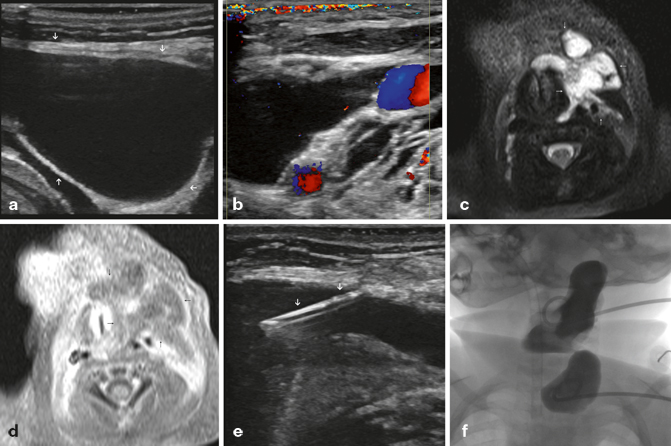
Fig. 22.3
A 3-month-old boy who presents with pain and swelling within his left neck. a Gray-scale ultrasound image shows large cyst within the soft tissues of the left neck consistent with a macrocystic lymphatic malformation (white arrow). b Color Flow Doppler ultrasound image of the large cyst within the soft tissues of the left neck demonstrates no vascular flow within the lesion. c Axial fat saturation T2-weighted MRI image of the neck, demonstrates an infiltrative area of an abnormal high signal within the soft tissues of the left neck (white arrows). d Corresponding axial fat saturation post contrast T1-weighted images shows that these areas have a peripheral rim of enhancement with a central area of low signal, consistent with a macrocystic lymphatic malformation (black arrows). e Gray-scale ultrasound image shows an angiocatheter entering the cyst (white arrows). f Spot fluoroscopic image shows two pigtail catheters in the cysts with contrast mixed with doxycycline
Doxycycline
Doxycycline is generally instilled into the cyst as a solution of 10 mg/ml. The dose of doxycycline can range from 150 to 1000 mg depending on the size of the lesion. In neonates, the maximum recommended dose at one time is 150 mg, because higher doses may cause hemolytic anemia, hypoglycemia, and metabolic acidosis [77]. Blood glucose is monitored 2 h post procedure in neonates. If the patient develops hypoglycemia, IV dextrose can be administered. A few small series have described a 93–100 % complete radiographic response for the treatment of macrocystic LMs [73, 78]. Lower response rates are noted for microcystic LMs.
Detergents
There is a limited literature on the used of detergents as sclerosant agents for LMs. One study described the use of 98 % ethanol and 3 % STS with a 100 % complete radiographic response rate [73]. A second series described the use of foamed STS for the treatment of 6 intraorbital LMs with a reduction in lesion size in all patients [79].
OK-432 (Picibanil)
OK-432 (Picibanil) is a lyophilized mixture of group A Streptococcus pyogenes, initially developed as an immunotherapeutic agent in the treatment of gastric and lung carcinoma with its first use as a sclerosant in the treatment of a LM described in a case report in 1987 [80]. Ok-432 is not approved in the USA. Direct injection of the solution into a LM has been shown to increase intralesional cytokine levels, namely tumor necrosis factor, interleukin 6 (Il-6), IL-8, interferon alpha, and vascular endothelial growth factor in patients with a response to treatment with Ok-432 [81, 82]. A review of the literature by Poldervaart et al. showed an 88 % excellent response rate for macrocystic lesions [83]. Microcystic lesions demonstrated an excellent response rates in 27 %, good in 33 %, and poor in 40 % of cases [83]. Adverse side effects were mild and consisted on fever, lethargy, and local inflammation. A phase II trial was conducted in the USA on 182 patients with LMs. In this trial, 94 % of the patient with macrocystic disease had a response to treatment, 63 % of the patients with mixed macrocystic/microcystic disease had a response to treatment, and 0 % of the patients with microcystic disease responded to treatment [66]. In two other series, complete response rates of 76 % and 83.5 % were noted [82, 84].
Alcohol Solution of Zein
Related posts:


Stay updated, free articles. Join our Telegram channel

Full access? Get Clinical Tree


