, Marilyn J. Siegel2, Tomasz Miszalski-Jamka3, 4 and Robert Pelberg1
(1)
The Christ Hospital Heart and Vascular Center of Greater Cincinnati, The Lindner Center for Research and Education, Cincinnati, OH, USA
(2)
Mallinckrodt Institute of Radiology, Washington University School of Medicine, St. Louis, Missouri, USA
(3)
Department of Clinical Radiology and Imaging Diagnostics, 4th Military Hospital, Wrocław, Poland
(4)
Center for Diagnosis Prevention and Telemedicine, John Paul II Hospital, Kraków, Poland
Abstract
The segmental, sequential, analytic approach was introduced by Van Praagh over 25 years ago [1–3]. This clinically useful approach is flexible and widely applicable to any imaging evaluation of congenital heart disease [4]. Using this logical approach, the morphologic features of the heart are determined sequentially by dividing the heart into three segments: visceroatrial situs or spatial arrangement (step 1), ventricular looping (step 2), and the spatial relationships of the great vessels (step 3). In step 4, the connections between the segments at the atrioventricular and ventriculoarterial levels are identified. Next, cardiac positioning is determined in step 5. Finally, step 6 involves the assessment for any associated malformation. See Fig. 5.1.
The segmental, sequential, analytic approach was introduced by Van Praagh over 25 years ago [1–3]. This clinically useful approach is flexible and widely applicable to any imaging evaluation of congenital heart disease [4]. Using this logical approach, the morphologic features of the heart are determined sequentially by dividing the heart into three segments: visceroatrial situs or spatial arrangement (step 1), ventricular looping (step 2), and the spatial relationships of the great vessels (step 3). In step 4, the connections between the segments at the atrioventricular and ventriculoarterial levels are identified. Next, cardiac positioning is determined in step 5. Finally, step 6 involves the assessment for any associated malformation. See Fig. 5.1.
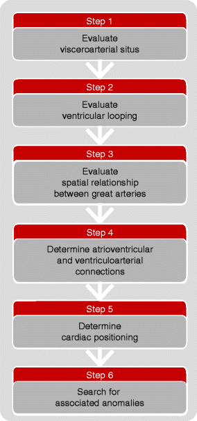
Fig. 5.1
A depiction of the approach to the segmental, sequential analysis of congenital heart disease. Letters are used to convey anatomic positioning of the three cardiac segments. The first three steps in the analysis involve the determination of atrial situs (step 1) (solitus, S, or inversus, I, or ambiguous, A), the ventricular looping pattern (step 2) (D- or L-), and the great artery spatial orientation (step 3) (solitus, S; inversus, I; D-TGA; L-TGA; A-TGA; D-MGA; L-MGA; A-MGA). Step 4 identifies the atrioventricular and ventriculoarterial connections. Step 5 determines cardiac positioning. Step 6 identifies associated malformations
This chapter describes the sequential steps required for accurate and complete analysis of complex congenital heart disease during a review of any imaging study. By following these steps, the reader of cardiac computed tomographic angiography studies, for example, will more likely end with an accurate interpretation. Using the Van Praagh nomenclature and initialing system, a segmental, sequential, analytic approach to the cardiac anatomy is applied.
5.1 Assessment of Cardiac Anatomy
5.1.1 Step 1: Determination of the Visceroatrial Situs
There are three possible arrangements of the atria (termed situs): solitus (S, -, -,) (the normal arrangement), inversus (I, -, -,) (the mirror image arrangement), and ambiguous (also called heterotaxy) (A, -, -,). Heterotaxy includes right isomerism [bilateral morphologic right atria and bronchial tree anatomy (two morphologic right lungs)] and left isomerism [bilateral morphologic left atria and bronchial tree anatomy (two morphologic left lungs)] (Fig. 5.3, panel c and d).
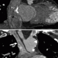
On imaging studies, anatomic differentiation of the morphologic right from left atrial chambers is usually based on the different morphology of the atrial appendages. The right atrial appendage is blunt and has a broad base communicating with the rest of the atrium. The left atrial appendage is narrower and has a more restricted junction with the smooth-walled left atrium and is more trabeculated than the right atrial appendage (Fig. 5.2). In addition, the inferior vena cava is associated with the right atrium.
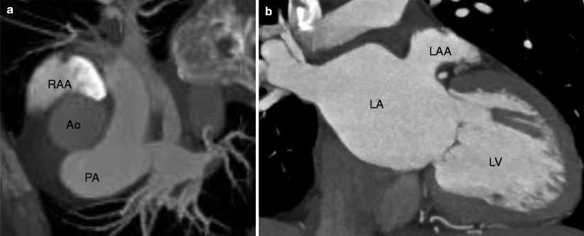
Fig. 5.2
Computed tomographic (CT) examples of the right (panel a) and left (panel b) atrial appendages identifying the right and left atria, respectively. The right atrial appendage has a larger, broader connection to the right atrium, and the left atrial appendage has more trabeculations and a narrower connection with the left atrium. RAA right atrial appendage, LAA left atrial appendage, RA right atrium, LA left atrium, RV right ventricle, LV left ventricle, PA pulmonary artery
If the appendages are not adequately distinguishable, then determination of atrial situs can be made based on the thoracic and visceral situs. The atrial situs almost always follows the thoracic and visceral situs. That is, if the left lung (bilobed) can be identified as being on the left and the right lung (trilobed) can be identified as being on the right and, concomitantly, the spleen and stomach are on the right and the liver on the left, then atrial situs solitus is most likely.
Inference can also be made from the spatial relationship among the abdominal great vessels and the spine (Fig. 5.3


Related posts:


Stay updated, free articles. Join our Telegram channel
Full access? Get Clinical Tree
