Shotgun Wounds
Daniel B. Nissman
FINDINGS
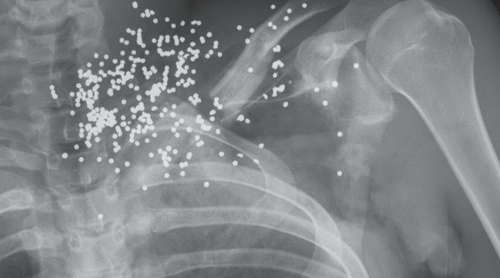
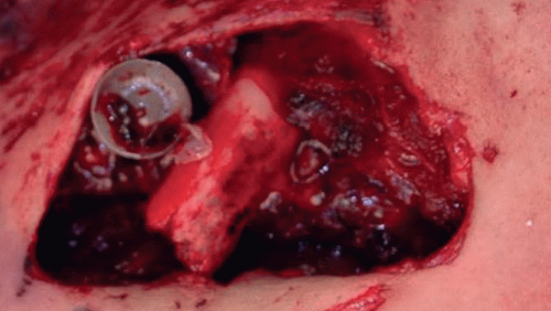
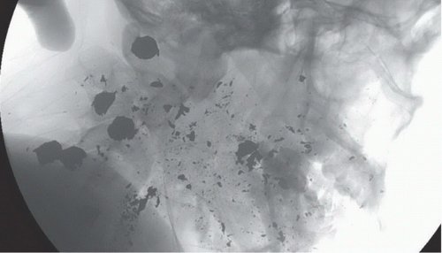
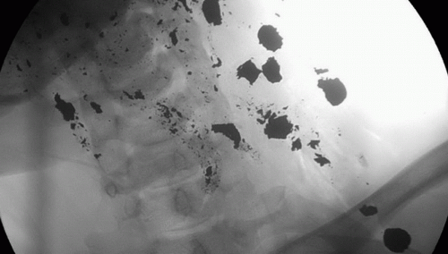
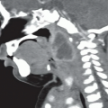
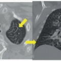
Postmortem AP radiograph of the left upper chest (Fig. 83A) reveals a comminuted midshaft clavicle fracture and numerous tiny, rounded metallic densities clustered about the clavicle, but mostly in the supraclavicular region. Note the relative size of the metallic densities in relation to the clavicular shaft; they are much smaller. Postmortem radiograph of the anterior upper chest centered on a large skin defect (Fig. 83B) reveals a piece of bone from the underlying comminuted clavicle fracture and a round, grayish appearing structure with central impression consistent with the wad from a shotgun shell. Lateral (Fig. 83C) and AP (Fig. 83D) fluoroscopic spot images from a postmortem examination of another patient reveal numerous large metallic densities clustered in the lateral left neck. The largest fragments are similar in size to the clavicular shaft (best seen in Fig. 83D). On the AP view much smaller fragments of metal density are noted along near-linear tracks adjacent to the larger metal density structures, much like a comet.
DIFFERENTIAL DIAGNOSIS
Low-energy hand gun injury, high-power hunting rifle injury, close-range shotgun injury, long-range shotgun injury.
DIAGNOSIS
Close-range shotgun blast with birdshot (Figs. 83A and B). Close-range shotgun blast with buckshot (Figs. 83C and D).
Related posts:


Stay updated, free articles. Join our Telegram channel

Full access? Get Clinical Tree


