Detecting signs of collapse within the lung is important in the diagnosis of lung disease. For us, it is also a good way to reinforce the anatomy. In general, the term collapse is used to describe markedly decreased volume of a lung, a lobe, or a segment. “Atelectasis” or “volume loss” is often used to describe less severe changes. The terms are fuzzy and interchangeable (hard to believe). First, let’s look at the patterns of collapse on x-ray and computed tomography (CT) and then possible mechanisms.
1
When a whole lung collapses, lung density increases, the volume diminishes, and adjacent structures move toward that lung. In Figure 8-1 , the left lung is consolidated and collapsed.
Figure 8-1
1
The trachea is ___________.
A.
midline
B.
left of midline
C.
right of midline
B.
left of midline
The heart has disappeared because ___________.
A.
there is Nocardia
B.
it shifted right
C.
it shifted left
C.
it shifted left
If the diaphragm were visible, it would be ___________.
A.
elevated
B.
depressed
C.
in a normal position
A.
elevated
2
The fissures that divide the lobes are formed by ___________.
A.
two parietal pleural layers
B.
two visceral pleural layers
C.
one visceral pleural layer
D.
one parietal pleural layer
2
B. two visceral pleural layers
3
Because fissures demarcate the boundaries of the lobes, the best sign of lobar collapse is shift of the fissures. Look at Figure 8-2 and decide which lobe has collapsed.
Figure 8-2A
Figure 8-2B
3
A.
right upper lobe
B.
right middle lobe
C.
right lower lobe
D.
left upper lobe
E.
left lower lobe
A.
___________
B.
___________
C.
___________
D.
___________
E.
___________
4
Let’s try this for real.
4
In Figure 8-3 , there is consolidation of the right ____________ lobe.
A.
upper
B.
middle
C.
lower
Figure 8-3
A.
upper
The sharp inferior margin is caused by the ____________ fissure.
A.
major
B.
minor
C.
superior accessory
D.
azygos
B.
minor
The fissure is ____________ in position.
A.
elevated
B.
depressed
C.
normal
A.
elevated
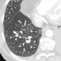
The accompanying CT scan shows collapse of the right upper lobe, and the arrow points to an endobronchial tumor obstructing the right upper lobe bronchus. Note absent air bronchogram.
The triangular density over the heart is the collapsed right __________ lobe.
A.
upper
B.
middle
C.
lower
B.
middle
In Figure 8-4B , there is a silhouette sign of the _________, caused by the right middle lobe collapse/consolidation.
A.
paratracheal area
B.
diaphragm
C.
right heart border
D.
left heart border
Figure 8-4B
C.
right heart border
6
The diagnosis of right middle lobe collapse on the frontal radiograph is often subtle. The diagnosis is often easier on the ___________ view.
A.
supine
B.
oblique
C.
frontal
D.
lateral
6
D. lateral
A triangular density, similar to right middle lobe collapse, may be present on the lateral, with collapse of the ______________.
A.
left upper lobe
B.
left lower lobe
C.
lingula
D.
left middle lobe
C.
lingula
Figure 8-5 shows collapse of two lobes on the right. The minor fissure is elevated, there is a silhouette sign of the upper mediastinum, and the trachea has shifted to the right because of right upper lobe collapse. There is a silhouette sign of the right diaphragm. The heart has moved to the right, indicating right lower lobe collapse. The right middle lobe remains aerated. We see the undersurface of the minor fissure and the right heart border because the right middle lobe is aerated.
On the frontal view ( Figure 8-6B ), there is a mass in the left hilum, and the left diaphragm is ________________.
A.
elevated
B.
depressed
C.
normal
Figure 8-6B
A.
elevated
This is a case of total collapse of the left ________________.
A.
upper lobe
B.
lower lobe
C.
upper lobe and lingula
D.
lingula
C.
upper lobe and lingula
The left upper lobe and lingula share a common bronchus. It is common for an endobronchial lesion (tumor, foreign body, mucus) to obstruct them together. In Figure 8-6A , the upper arrow is at the level of the upper lobe, and the lower arrow is at the level of the lingula.
8
Similarly, the bronchus intermedius on the right supplies the right _________ and _________ lobes.
A.
upper
B.
middle
C.
azygos
D.
lower
8
B. middle and D. lower
Figure 8-7 shows dense consolidation at the right base. The minor fissure is ________________.
A.
elevated
B.
depressed
C.
normal
Figure 8-7
B.
depressed
There are silhouette signs of the right heart and diaphragm.
The right ________________ lobe(s) is (are) collapsed.
A.
upper
B.
middle
C.
lower
B.
middle and C. lower
9
On the left, the _____________ share a common bronchus.
A.
upper lobe
B.
lingula
C.
lower lobe
9
A. upper lobe and B. lingula
On the right, the middle and lower lobes share a common bronchus, called the _____________.
A.
azygos bronchus
B.
right middle lobe bronchus
C.
right lower lobe bronchus
D.
bronchus intermedius
D.
bronchus intermedius
A complete obstruction of either bronchus causes collapse of _____________ lobe(s).
A.
one
B.
two
C.
three
B.
two
Movement of the fissures is the most reliable sign of lobar collapse. Crowding of pulmonary vessels or bronchi or movement of parenchymal landmarks (e.g., nodules, granulomas, surgical clips) also can indicate volume loss.
10
If a lobe or segment is atelectatic, but still contains some air, the vascular markings would be visible, but in a _____________ volume.
A.
smaller
B.
larger
C.
similar
10
A. smaller
If the lung is atelectatic and consolidated, the air bronchogram sign might show us the bronchi. In either case, the vessels or bronchi would appear _____________.
A.
further apart
B.
crowded together
C.
invisible
B.
crowded together
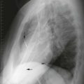
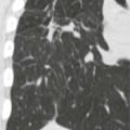
Figure 8-8 shows crowded air bronchograms in left lower lobe collapse (arrows). The collapsed lung itself is difficult to see behind the heart. There is also a silhouette sign of the left diaphragm.
11
In Figure 8-9A , there is a subtle nodule in the right upper lobe. In Figure 8-9B , after a needle biopsy, the nodule position is _____________.
A.
unchanged
B.
more lateral
C.
more medial
Only gold members can continue reading. Log In or Register to continue