Reused with permission. From Austin JH, Muller NL, Friedman PJ, et al. Glossary of Terms for CT of the Lungs: Recommendations of the Nomenclature Committee of the Fleischner Society Radiology 1996, Aug; 200(2): 327-331© RSNA 1996
John H. M. Austin, MD *
Nestor L. Müller, MD, PhD
Paul J. Friedman, MD
David M. Hansell, MB
David P. Naidich, MD
Martine Remy-Jardin, MD
W. Richard Webb, MD
Elias A. Zerhouni, MD
In 1991, Drs. Webb, Müller, and Naidich offered an introductory glossary of the vocabulary of thin-section computed tomography (CT) in their book, High-Resolution CT of the Lung, and updated that glossary in 1993. The present committee, by using those contributions as a base, now offers a considerably expanded glossary. Our intent is to list, define, and provide references for the main terms used in chest CT that are specific to the lungs. “HRCT” is used to indicate high-resolution computed tomography.
Use of words is inherently controversial. We are pleased to invite readers to offer improvements to our definitions.
Index terms: Computed tomography Lung, CT
* From the Department of Radiology, Columbia-Presbyterian Medical Center, 622 W 168th St., New York, NY 10032-3784 (J.H.M.A.); Department of Radiology, University of British Columbia and Vancouver Hospital and Health Sciences Centre, Canada (N.L.M.); Department of Radiology, University of California San Diego, School of Medicine (P.J.F.); Department of Radiology, Royal Brompton National Heart and Lung Hospital, London, England (D.M.H.); Department of Radiology, New York University Medical Center, NY (D.P.N.); Department of Radiology, Hopital Calmette, Lille, France (M.R.-J.); Department of Radiology, University of California San Francisco, School of Medicine (W.R.W.); Department of Radiology, The Johns Hopkins University School of Medicine, Baltimore, Md (E.A.Z.). Received November 10, 1995; revision requested February 12, 1996; revision received March 18; accepted April 26. Address reprint requests to J.H.M.A. ©RSNA, 1996
Glossary
air crescent: Air in a crescentic shape in a nodule or mass, in which the air separates the outer wall of the lesion from an inner sequestrum, which most commonly is a fungus ball of Aspergillus species.
air trapping:
- 1.
Pathophysiology . The retention of excess gas (“air”) in all or part of the lung, especially during expiration, either as a result of complete or partial airway obstruction or as a result of local abnormalities in pulmonary compliance. Although not in common usage, the term “gas trapping” is more accurate.
- 2.
CT . Decreased attenuation of pulmonary parenchyma, especially manifest as less than normal increase in attenuation during expiration. To be differentiated from the decreased attenuation of hypoperfusion secondary to locally increased pulmonary arterial resistance.
- 1.
architectural distortion: A manifestation of lung disease in which bronchi, pulmonary vessels, a fissure or fissures, or septa of secondary pulmonary lobules are abnormally displaced.
band: See “parenchymal band.”
beaded septum sign: Irregular septal thickening that suggests the appearance of a row of beads; usually a sign of lymphangitic carcinomatosis but may also occur rarely in sarcoidosis. Because the thickening usually is more irregular than beaded, the term “irregular septal thicken” generally is preferred.
bronchiectasis:
- 1.
Pathology . Irreversible dilatation of a bronchus or bronchi, often with thickening of the bronchial wall. When mild, the dilatation is cylindric (ie, normal bronchial tapering is absent). When more severe, the dilatation is saccular, and irregular constrictions may be present. When very severe, the bronchi may be markedly dilated, especially distally. See also “traction bronchiectasis.”
- 2.
CT . Bronchial dilatation, often with thickening of the wall.
- 1.
bronchiolectasis:
- 1.
Pathology. Dilatation of a bronchiole or bronchioles, often with thickening of the bronchiolar wall.
- 2.
CT . Bronchiolar dilatation. See also “traction bronchiolectasis.”
- 1.
bulla:
- 1.
Pathology . A sharply demarcated, dilated air space that measures 1 cm or more in diameter and possesses a thin epithelialized wall, which is usually no greater than 1 mm in thickness. See also “bullous emphysema,” “emphysema,” and “paraseptal emphysema.”
- 2.
CT . A round, focal air space, 1 cm or more in diameter, demarcated by a thin wall; usually multiple or associated with other signs of pulmonary emphysema.
- 1.
bullous emphysema: Emphysema characterized by the presence of bullae. See “emphysema.”
centriacinar emphysema: See “centrilobular emphysema.”
centrilobular: Referring to the region of the bronchioloartenolar pulmonary lobule. See “centrilobular structures.”
centrilobular emphysema:
- 1.
Pathology . Emphysema that is characterized by destroyed centrilobular alveolar septa and enlargement of respiratory bronchioles. Usually in the upper lung zones of cigarette smokers.
- 2.
CT. Centrilobular decreased attenuation, usually without visible walls, of nonuniform distribution, and predominantly located in upper lung zones. Synonym: centriacinar emphysema.
- 1.
centrilobular structures:
- 1.
Anatomy . The central tubular structures in a secondary pulmonary lobule (ie, the centrilobular artery and bronchiole).
- 2.
CT . The pulmonary artery and its immediate branches in a secondary lobule; these arteries measure approximately 1 mm and 0.5-0.7 mm in diameter, respectively; HRCT depicts these vessels. However, a normal bronchiole supplying a secondary lobule has a wall thickness of approximately 0.15 mm, which is beyond the resolution of HRCT. Therefore, normal airways in secondary pulmonary lobules are not detected at CT examination.
- 1.
consolidation:
- 1.
Pathology . Transudate, exudate, or tissue replacing alveolar air.
- 2.
CT. Homogeneous increase in pulmonary parenchymal attenuation that obscures the margins of vessels and airway walls. An air bronchogram may be present.
- 1.
core structures: See “centrilobular structures.”
cyst:
- 1.
Pathology . A round, circumscribed space that is surrounded by an epithelial or fibrous wall of thickness, which may be uniform or varied, and that in the lung usually contains air but may contain liquid, semisolid, or solid material.
- 2.
CT. A round, parenchymal space with a well-defined wall; usually air-containing when in the lung but without associated pulmonary emphysema; commonly used to describe enlarged air spaces in end-stage fibrosis of idiopathic pulmonary fibrosis and sarcoidosis, and also in Langerhans cell histiocytosis and lymphangiomyomatosis. See “bulla,” “cystic air space,” and “honeycomb cysts.”
- 1.
cystic air space: Enlarged unit of peripheral air-containing lung, surrounded by a wall of variable thickness, which may be thin as in lymphangiomyomatosis, or may be thick as in idiopathic pulmonary fibrosis. See “bulla,” “cyst,” and “honeycomb cysts.”
dependent increased attenuation: See “dependent opacity.”
dependent opacity: Subpleural increased attenuation in dependent lung. The increased attenuation disappears when the region of lung is nondependent. May also appear as a subpleural line.
distal acinar emphysema:
- 1.
Pathology . Emphysema characterized by predominant involvement of alveolar ducts and sacs, characteristically in subpleural lung and adjacent to interlobular septa and vessels.
- 2.
CT. Emphysema characterized by subpleural regions of low attenuation or bullae separated by intact interlobular septa. Synonym: paraseptal emphysema.
- 1.
distortion: See “architectural distortion.”
emphysema:
- 1.
Pathology . Permanently enlarged air spaces distal to the terminal bronchiole, accompanied by destroyed alveolar walls. Absence of “obvious fibrosis” historically has been regarded as an additional criterion, but the validity of that criterion recently has been called into question.
- 2.
CT. Focal region or regions of low attenuation, usually without visible walls, resulting from actual or perceived enlarged air spaces and destroyed alveolar walls. May be associated with air trapping. See also “bulla,” “bullous emphysema,” “centrilobular emphysema,” “cyst,” “cystic air space,” “distal acinar emphysema,” “panlobular emphysema,” and “paraseptal emphysema.”
- 1.
fungus ball: A masslike collection of intertwined hyphae, usually Aspergillus species, matted together by mucus, fibrin, and cellular debris and colonizing a pulmonary cavity caused by prior disease (e.g., sarcoidosis). May move to a dependent location when the patient changes position. At CT, may show a “spongework” pattern, including foci of high attenuation. Synonym: mycetoma.
gas trapping: See “air trapping.”
ground-glass attenuation: See “ground-glass opacity.”
ground-glass opacity: Hazy increased attenuation of lung, but with preservation of bronchial and vascular margins; caused by partial filling of air spaces, interstitial thickening, partial collapse of alveoli, normal expiration, or increased capillary blood volume. Not to be confused with “consolidation,” in which bronchovascular margins are obscured. May be associated with an air bronchogram.
halo sign: Ground-glass opacity surrounding the circumference of a nodule or mass. May be a sign of invasive aspergillosis or hemorrhage of various causes.
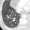
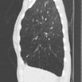
honeycomb cysts: Cystic air spaces, usually of comparable diameter and on the order of 0.3-1.0 cm in diameter, formed by the honeycombing of interstitial pulmonary fibrosis.
honeycombing:
- 1.
Pathology . Destroyed, fibrotic, and cystic lung, representing complete loss of acinar and bronchiolar architecture as the end stage of fibrosing lung disease.
- 2.
CT . Clustered cystic air spaces, usually of comparable diameters on the order of 0.3-1.0 cm but as much as 2.5 cm, usually subpleural and characterized by well-defined walls, which are often thick. A CT feature of diffuse pulmonary fibrosis. A diagnostic pitfall is that, in the presence emphysema, air-space consolidation can mimic this appearance.
- 1.
interlobular septal thickening: See “septal line.”
intralobular lines: Fine linear opacities present in a lobule when the intralobular interstitium is thickened. When numerous, they may appear as a fine reticular pattern.
irregular linear opacity: Any linear opacity of irregular thickness of 1-3 mm, distinct from interlobular septa, bronchovascular bundles, and nodular opacities. May be intralobular or extend through several adjacent secondary lobules.
linear opacity: An elongated, thin line of soft-tissue attenuation. Rarely, calcification or foreign material may increase the attenuation. See also “irregular linear opacity” and “subpleural line.”
lobular core structures: See “centrilobular structures.”
lobule: See “secondary pulmonary lobule.”
micronodule: Discrete, small, round, focal opacity of at least soft-tissue attenuation and with a diameter no greater than 7 mm. Some authors have limited use of this term to a diameter of less than 5 mm or less than 3 mm. Other authors simply use the term “small nodule.” See “nodule.”
midlung window: A midlung region, characterized by the absence of large blood vessels and by a paucity of small blood vessels that corresponds to the minor fissure and adjacent peripheral lung.
mosaic oligemia: See “mosaic perfusion.”
mosaic perfusion: A patchwork of regions of varied attenuation, interpreted as secondary to regional differences in perfusion. A more inclusive term than the originally described “mosaic oligemia.” Air trapping secondary to bronchial or bronchiolar obstruction may also produce focal zones of decreased attenuation, an appearance that can be enhanced by using expiratory CT.
mycetoma: See “fungus ball.”
nodule:
- 1.
Pathology . Small, approximately spherical, circumscribed focus of abnormal tissue.
- 2.
Radiology . Round opacity, at least moderately well marginated and no greater than 3 cm in maximum diameter. Some authors use the modifier “small” if the maximum diameter of the opacity is less than 1 cm. See also “micronodule.”
- 1.
opacification: See “parenchymal opacification.”
panacinar emphysema: See “panlobular emphysema.”
panlobular emphysema:
- 1.
Pathology. Emphysema that involves, more or less uniformly, all portions of the secondary lobules. It tends to predominate in the lower lobes and is the form of emphysema associated with hereditary α1-protease inhibitor (α1-antitrypsin) deficiency.
- 2.
CT . Emphysema that tends to show rather uniformly decreased parenchymal attenuation and a paucity of vessels. Severe panlobular emphysema may be indistinguishable from severe centrilobular emphysema, except on the basis of zonal distribution. Synonym: panacinar emphysema.
- 1.
paraseptal emphysema: See “distal acinar emphysema.”
parenchymal band: Elongated opacity, usually several millimeters wide and up to about 5 cm long, often extending to the pleura, which may be thickened and retracted at the site of contact. Originally described in asbestosis but also a sign of focal fibrosis of non-specific cause.
parenchymal opacification: Increase in pulmonary attenuation that may or may not obscure the margins of vessels and airway walls. “Consolidation” indicates that definition of these margins (excepting air bronchograms) is lost, whereas “ground-glass opacity” indicates a lesser increase in attenuation, in which definition of the margins is preserved. Whenever possible, use of the more specific terms “consolidation” or “ground-glass opacity” is preferred.
peripheral: Referring to pulmonary structures within 1-2 cm of any visceral pleural surface. See also “subpleural.”
pseudoplaque: An irregular band of peripheral pulmonary opacity adjacent to visceral pleura that simulates the appearance of a pleural plaque and is formed by the coalescence of small nodules (e.g., in coal-worker’s pneumoconiosis).
reticular pattern: See “reticulation.”
reticulation: Innumerable, interlacing line shadows that suggest a mesh. A descriptive term usually associated with interstitial lung diseases. May be fine, intermediate, or coarse. Synonym: reticular pattern.
secondary pulmonary lobule:
- 1.
Anatomy . The smallest unit of lung surrounded by connective tissue septa, according to Miller and Heitzman et al. These septa, known as interlobular septa, are best developed in the periphery of the anterior, lateral, and juxtamediastinal regions of the upper and middle lobes and in the periphery of the anterior and diaphragmatic regions of the lower lobes. The septa tend to be incompletely developed or absent elsewhere in the lungs. Miller’s lobule ranges in size from 0.5 to 3.0 cm and may contain 3-20 acini.
- 2.
Anatomy. The unit of lung subtended by any bronchiole that gives off three to five terminal bronchioles, according to Reid. Connective tissue septa are not part of this definition. A small Miller’s lobule (0.5 cm) corresponds to a Reid’s lobule.
- 3.
CT. Miller’s lobule is the secondary lobule that is identified with CT. See also “centrilobular structures.”
- 1.
septal line: Thin linear opacity that corresponds to an interlobular septum; to be distinguished from centrilobular structures. See “septal thickening.”
septal thickening: Abnormal widening of an interlobular septum or septa, usually caused by edema, cellular infiltration, or fibrosis. May be smooth, irregular, or nodular. See also “beaded septum sign.”
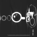
signet-ring sign: A ring of opacity (usually representing a dilated, thick-walled bronchus) in association with a smaller, round, soft-tissue opacity (the adjacent pulmonary artery or, rarely, dilated bronchial artery) suggesting a “signet ring.” Usually this finding indicates bronchiectasis, but it may also occur in multifocal bronchioloalveolar carcinoma and metastatic adenocarcinoma.
subpleural: Referring to pulmonary structures that are next to or near visceral pleura.
subpleural line: A thin curvilinear opacity, a few millimeters or less in thickness, usually less than 1 cm from the pleural surface and paralleling the pleura. A nonspecific indicator of atelectasis, edema, fibrosis, or inflammation. See also “irregular linear opacity.”
traction bronchiectasis: Bronchial dilatation, which is commonly irregular, in association with juxtabronchial opacification that is interpreted as representing retractile pulmonary fibrosis.
traction bronchiolectasis: Bronchiolar dilatation in association with peribronchiolar opacification that is interpreted as representing retractile pulmonary fibrosis.
tree-in-bud sign: Nodular dilatation of centrilobular branching structures that resembles a budding tree and represents exudative bronchiolar dilatation (e.g., in panbronchiolitis or endobronchial spread of active pulmonary tuberculosis.
Related posts:

Stay updated, free articles. Join our Telegram channel

Full access? Get Clinical Tree


