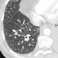
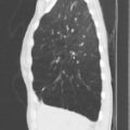
Ground Glass Opacification (Synonyms: foggy, hazy, and semiopaque )


Comments
This is like looking at the anatomy through a frosted shower door or glass. The lung is an intermediate shade of gray but the pulmonary vessels are visible within the grey areas. The diminished aeration may be due to (1) decreased air in the alveoli caused by partial alveoli filling, (2) decreased air in the alveoli caused by thickened interstitium encroaching on the alveoli, or (3) decreased air in the alveoli due to hypoventilation and atelectasis.
Causes
atelectasis
aspiration pneumonitis
infection, such as pneumocystis
edema, acute respiratory distress syndrome (ARDS)
pulmonary hemorrhage
idiopathic (e.g., desquamative interstitial pneumonitis, chronic organizing pneumonia)
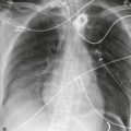
Reticular (Synonyms: linear and irregular )


Comments
Acute or chronic thickening of the interlobular septa or the bronchovascular bundles cause linear or lacelike thickening. This may be smooth or irregular.
Causes
edema (Kerley lines)
lymphangitic tumor
sarcoidosis
Langerhans histiocytosis (eosinophilic granuloma)
fibrosis (any cause)
Micro nodules (Synonym: miliary nodules )