4] who recognized that a suitable software package in conjunction with a diagnostic CT scanner could be developed that would emulate a conventional radiotherapy simulator. In the early 1980s, the use of CT scanners in treatment planning amounted to providing axial images that could be transferred to the TPS. The process of plan optimization and verifying the set-up before treatment was purely a role for the TPS and the physical simulator with no significant manipulation of the 3D CT data to augment this process. At this time, the use of the Beam’s Eye View (BEV) allowed the visualization of a beam shape relative to the target contour, bony anatomy and other contoured organs. The BEV gave the planner the ability to optimize beam positions, shapes and orientation in the same manner as could be achieved radiographically on the simulator.
In 1983, Goitein et al [
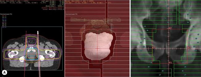
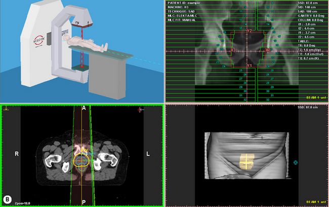
The automatic registration of the BEV and DRR have provided powerful tools to enable the planner to achieve on the TPS what was available on the simulator but with the added benefits of CT visualization (see


Figure 9.5 (A) Beams Eye View (BEV) on a Treatment Planning System with Digitally Reconstructed Radiograph (DRR) on Virtual Simulator; (B) Screen shot of Virtual Simulator with surface rendering and transverse slice.
Stay updated, free articles. Join our Telegram channel

Full access? Get Clinical Tree


