Abstract
The skin and subcutaneous tissues are common sites of inflammatory lesions which must be distinguished from malignancy. Visual inspection of FDG-avid skin lesions is often needed to make this distinction. Likewise, FDG avid lesions in breast tissue may be benign or malignant. Incidentally detected FDG-avid breast lesions should be evaluated by mammogram and/or ultrasound.
Keywords
FDG, PET/CT, skin, breast, breast cancer
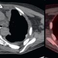
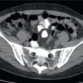
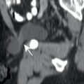
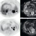
The skin and subcutaneous tissues are common sites of inflammatory lesions such as acne, sebaceous cysts, warts, and pilonidal cysts ( Fig. 5.1 ). These benign etiologies may produce abnormalities on 18F-fluorodeoxyglucose positron emission tomography/computed tomography (FDG PET/CT), or both. However, the skin is also the site of primary malignancies such as melanoma, squamous cell carcinoma, and Kaposi sarcoma ( Fig. 5.2 ), as well as lymphoma ( Fig. 5.3 ) or metastases ( Fig. 5.4 ). It is usually not possible to distinguish these benign and malignant lesions on whole-body FDG PET/CT. Fortunately, the skin allows for direct inspection of abnormalities. Thus often the best method for further evaluation of abnormality seen on FDG PET/CT is the suggestion to perform a physical examination to correlate with imaging findings. Usually the patient is no longer available to the radiologist when a skin or subcutaneous lesion is identified on PET/CT images. In clinics where the patient is available to the interpreting radiologist, this is one abnormality where the radiologist can perform their own physical examination. A PET/CT report which describes the radiologist’s physical examination findings is sure to raise eyebrows in your institution.


In patients with known breast cancer, the FDG-avid soft tissue lesions within the breast probably represent the primary breast malignancy ( Fig. 5.5 ). However, in patients without known breast malignancy, FDG PET/CT is not sensitive for the detection or diagnosis of breast lesions. Due to physical examination and mammographic screening, many breast cancers are small, early stage lesions at diagnosis, which are often not visualized by either whole-body FDG PET or CT, even though they are readily apparent on dedicated breast imaging such as mammography, ultrasound, or breast magnetic resonance (MR) ( Fig. 5.6 ).


