Skull


Skull


Edward Yang

Sara O. Vargas

Tina Young Poussaint


INTRODUCTION

Suspected skull disorders are common indications for imaging in the pediatric age group, being easily identified by caregivers and physicians because of their superficial location. As an osseous structure, the skull manifests many of the pathologies seen elsewhere in the skeletal system, but it does so in the setting of dynamic changes of development and maturation of the skull itself as well as the brain that it protects.

This chapter reviews the radiographic anatomy and disorders affecting the skull as well as its connective tissue covering, the scalp, in the pediatric population. The chapter begins by considering the various modalities for performing an imaging examination of the pediatric skull. Next, normal imaging anatomy and skull development are discussed. Then, an overview is provided of the most commonly encountered diseases of the pediatric skull, including disorders of skull shape and integrity, infectious disorders, diffuse bone diseases, neoplasms affecting the skull and scalp, and features of skull trauma unique to the pediatric population.


IMAGING TECHNIQUES


Radiography

Radiography is the oldest imaging technique available for evaluating the skull and remains widely used because of its low cost, wide availability, and suitability in unsedated patients. In addition, it is associated with relatively low radiation dose compared to computed tomography (CT). Current indications include detection of diffuse bone abnormalities as part of a skeletal survey (e.g., metastatic disease, metabolic bone disease), screening for nonaccidental trauma (child abuse) in a neurologically intact child <2 years, and screening for craniosynostosis. Skull radiographs are also commonly used to assess surgical implants (e.g., shunts, programmable shunt valves, and cochlear implants) and to exclude the presence of radiopaque foreign bodies.

Standard skull radiography technique consists of frontal (usually posteroanterior [PA]), lateral, and Townes projections (Fig. 1.1). Whereas the petrous apices project over the central orbits in the PA projection, they are above the orbits in the Townes view because of angulation used for better visualization of the occipital bone and foramen magnum. An optional view opening up the frontal bone is the Caldwell projection where the petrous apices lie below the orbits. An age-specific dose is delivered with collimation to cover the osseous structures and overlying scalp. Typical delivered skin dose is ˜1 mGy, and there is an estimated effective dose of ˜0.02 mSv under these conditions.1


Ultrasound

Sound waves penetrate osseous structures poorly and therefore ultrasound is not a widely used imaging modality for the skull in most institutions. However, the availability of ultrasound at the point of care and the lack of radiation exposure have prompted the use of ultrasound for selected indications in young (<2 years old) patients who have little hair and relatively sonolucent osseous structures. Specifically, ultrasound has been used to successfully detect skull fractures and sutural synostosis.2,3,4,5 Additionally, ultrasound has long been a firstline tool for evaluating scalp masses.6,7

Ultrasound of the skull uses high-frequency (typically >8 MHz) linear transducers coupled with transducing gel. Adjustments are performed to reduce the depth and focus to accommodate the superficial structures being imaged. Typically, stand-off pads are not required. For soft tissue or
vascular scalp lesions, color and duplex Doppler are added to demonstrate lesional vascularity and pulsatility of blood flow (i.e., arterial versus venous).


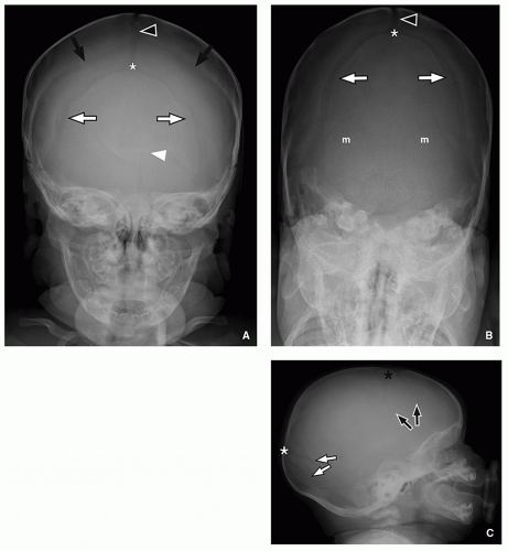
FIGURE 1.1 Skull radiographs of a normal 3-monthold boy: posteroanterior (PA) (A), Townes (B), and lateral (C) projections. Black arrows indicate coronal sutures, black arrowhead the sagittal suture, white arrows the lambdoid sutures, white arrowhead the metopic suture, white asterisk the posterior fontanelle, black asterisk the anterior fontanelle, and “m” the mendosal sutures.


Computed Tomography

As a high-resolution, cross-sectional technique with excellent osseous detail and soft tissue contrast, computed tomography (CT) is the current reference standard for indications such as head trauma, craniosynostosis, and characterization of focal skull lesions. Although some authors have proposed algorithms that omit CT for single-suture synostosis,8,9,10 CT has superior sensitivity/specificity for detecting synostosis as it can detect even small areas of osseous bridging that may elude radiography.11,12,13 There are also data to suggest that CT becomes increasingly cost-effective for individuals with a high pretest likelihood of synostosis, particularly involving multiple sutures. Regardless of diagnostic strategy for abnormal head shape, common clinical practice is to obtain a head CT with three-dimensional (3D) surface rendering for cases that are scheduled for surgery as the surface model assists procedural planning.11,14 For reasons discussed later (see “Fractures”), the American College of Radiology Appropriateness Criteria endorses an approach that integrates head CT when imaging is pursued for abusive or nonabusive head trauma.15,16

Acquisition of a head CT depends on both the precise hardware available as well as the indication. For most modern multidetector CT instruments, axial acquisition of the head is followed by generation of both submillimeter (isotropic) and 3- to 5-mm-thick data using soft tissue (brain) and bone kernels. The submillimeter data can then be used to
create multiplanar reformatted images and surface rendered data, the latter particularly helpful in delineating complex 3D relationships of skull lesions. In our institution, an agespecific dosage scheme17 compliant with recommendations from both the Society for Pediatric Radiology Image Gently campaign and the ACR CT Accreditation Requirements is used,18,19 the latter limiting the CT dose index (CTDIvol) for a 1-year-old to 40 mGy compared to 80 mGy for an adult. For pediatric patients undergoing evaluation for craniofacial dysmorphism, the field of view is extended below the mandible to capture any facial bone abnormalities, and the dose is reduced to 100 kV and 50 mAs, resulting in a CTDI <5 mGy. The trade-off for this reduced dose is poorer parenchymal detail. Even with these attempts to reduce dose, it is worth noting that a standard head CT still carries an estimated dose that is 20 times a single projection skull radiograph.20


Magnetic Resonance Imaging

Magnetic resonance imaging (MRI) has several advantages for imaging of the skull and overlying scalp including superior soft tissue contrast and the ability to suppress fat signal within the skull or scalp. For these reasons, MRI is often used to further characterize soft tissue masses in the scalp and discrete osseous lesions (e.g., primary bone tumors, metastases).21,22,23,24 It also easily depicts communication of scalp/skull lesions with the intracranial contents, a common concern when the differential diagnosis includes lesions such as cephaloceles, sinus pericranii, and dermoid cysts.

The optimal MR protocol for evaluation of a scalp or cranial vault lesion includes at least two planes of fat-suppressed T2-weighted MR imaging (usually, short tau inversion recovery [STIR]) and two planes of contrast-enhanced T1-weighted MR imaging with fat suppression. Advanced imaging techniques such as diffusion-weighted imaging have also been applied to the skull to assess for pathology such as metastatic disease and osteomyelitis.25,26 In combination with standard brain MRI sequences, these additional sequences make intrinsic marrow signal abnormalities, focal osseous lesions, and soft tissue masses of the scalp conspicuous as the background signal from fat is suppressed. However, erosion of the skull and skull fractures remain poorly seen compared to CT because of the comparatively low spatial resolution of MR.27,28 New MRI techniques are attempting to address these limitations but are not yet widely used outside of research settings.29,30


Nuclear Medicine

Positron and gamma ray emitting radiotracers are rarely used specifically to detect skull pathology. In the case of skull fractures (inflicted or accidental), it is widely accepted that skull fractures do not reliably appear on technetium-based bone scans because of poor callus formation though bone scintigraphy remains commonly performed for detection of fractures elsewhere.31,32 Whether introduction of 18F-fluorine PET improves sensitivity awaits further investigation.33 For infections of the skull, there is adult literature that suggests gallium and technetium radiotracers may have some utility particularly for skull base infections,34,35,36 but MRI can provide similar information while simultaneously evaluating for intracranial pathology. For oncologic indications, several radiotracers remain in wide use for screening of metastatic disease and can therefore suggest skull involvement. These include staging for tumors common to the cranial vault such as Langerhans cell histiocytosis (technetium 99m methylene diphosphonate [Tc99m-MDP] and 18F-fluorodeoxyglucose [FDG])37,38 and neuroblastoma (Tc99m-MDP, 123I-metaiodobenzyguanidine [MIBG], and FDG).39,40


NORMAL ANATOMY


Major Sutures and Fontanelles

Sutures are fibrous articulations between bones of the cranial vault that develop by intramembranous ossification. The skull base (sphenoid, ethmoid, nonsquamous temporal bone, and occipital bone below the superior nuchal line) develop with enchondral ossification, and therefore the articulations of the skull base ossification centers are called synchondroses.41,42,43,44 The major sutures of the cranial vault include the sagittal suture separating the paired parietal bones, the metopic suture separating the two halves of the frontal bone, the coronal suture separating the frontal and parietal bones, and the lambdoid suture separating the occipital and parietal bones (Figs. 1.1 and 1.2).

Major openings present at birth include the anterior fontanelle at the junction of the coronal/sagittal sutures (called the bregma when closed) and the posterior fontanelle at the junction of the lambdoid sutures (called the lambda when closed).


Accessory/Minor Sutures

A number of minor sutures are seen in the vicinity of the squamous temporal bone, including the sphenotemporal (sphenosquamous) suture anteriorly, temporoparietal (squamous) suture superiorly, and the occipitomastoid suture posteriorly (Fig. 1.2).45 The H-shaped junction where the sphenoparietal suture meets the coronal/frontosphenoid sutures anteriorly and the sphenotemporal/temporoparietal sutures posteriorly is called the pterion. The complexity of occipital bone development (at least eight ossification centers) gives rise to a number of transient sutures in this region (Fig. 1.3). Common transient sutures include midline superior and inferior occipital fissures at the lambda and foramen magnum, respectively; mendosal sutures including forms dividing the squamous occipital bone transversely; and paired anterior/posterior intraoccipital synchondroses that straddle the anterior and posterior extent of the foramen magnum at the skull base.46,47,48


Normal Development and Timing of Suture Closure

The initial ossification of the skull begins at a few months of gestation and is largely complete by term delivery.49 The
sutures and skull base synchondroses are patent at birth and then begin to close at variable rates. However, two useful rules of thumb are that no major suture should close in the first year of life and no suture should undergo mature fusion in childhood except for the metopic suture, which normally closes at 3 to 9 months.44,50,51,52,53,54 As the patient ages, the sutures become more serrated at their outer table though they remain smooth along the inner table.55,56 For cases where there is subjective narrowing, normative neonatal data have been published for selected sutures based on CT data.57 Important synchondrosis milestones include closure of frontosphenoid and intersphenoid synchondroses by 1 to 2 years of age and of the sphenoccipital (clival) synchondroses during adolescence.58 Closure of synchondroses and sutures within the occipital bone follow a more complicated sequence. The synchondroses at the foramen magnum typically close within the first few months of life though other sutures including mendosal sutures and inferior occipital fissure may persist through the first few years of life47,48,58,59; variants where these sutures/synchondroses persist into adulthood are occasionally encountered. The anterior and posterolateral fontanelles typically close by the end of the 2nd year. The posterior fontanelle closes much earlier, typically by 3 to 6 months.60


FIGURE 1.2 Three-dimensional surface renderings of a head CT from a normal 3-month-old boy evaluated for craniosynostosis: frontal (A), lateral (B), posterior (C), and superior (D) views. Annotations as for Figure 1.1 with additional markings of pterion with a circle, squamous (temporoparietal) suture with “s,” sphenotemporal suture with white dashed arrow, frontosphenoid suture with black dashed arrow, occipitomastoid suture with “o,” and posterior intraoccipital synchondrosis with “p.”

Deviations from normal sutural closing (apparent sutural widening) are present in conditions of poor bone
mineralization (osteogenesis imperfecta, rickets, hypothyroidism) and can be misinterpreted as sutural diastasis from elevated intracranial pressure. Other reported causes of apparent widening of normal sutural width include recovery from chronic malnutrition, in utero renin-angiotensin system disruption (Fig. 1.4), achondroplasia, trisomy 21, and treatment with prostaglandins for prematurity.61,62,63,64,65


FIGURE 1.3 Transient sutures of the occipital bone visualized on a three-dimensional surface rendering of a 1-monthold boy’s head CT (posterior view). Markings are as for Figures 1.1 and 1.2 but additional sutures marked include “so”, “m”, and “io” for superior occipital, paired mendosal sutures, and inferior occipital sutures, respectively.


FIGURE 1.4 Markedly widened cranial sutures (hypocalvarium) in a 1-day-old girl with renal tubular agenesis, pulmonary hypoplasia, and other skeletal anomalies suggesting in utero exposure to renin-angiotensin system blockers or an inborn error in the renin-angiotensin system. Reflection of the scalp at autopsy shows only a thin membrane covering much of the cerebrum. In the right panel, the margins of the fontanelles are outlined in black ink.


SPECTRUM OF SKULL DISORDERS


Congenital and Developmental Anomalies


Craniosynostosis

Craniosynostosis refers to premature closure of the cranial sutures (segmental or total) with resultant deformity. Craniosynostoses are usually classified as primary or secondary, secondary forms representing the consequence of an identifiable cause unrelated to suture development, such as metabolic bone disease, bone dysplasia, or loss of intracranial volume (e.g., shunting, brain injury). The primary craniosynostoses are further divided into single versus multiple suture forms and syndromic versus nonsyndromic (isolated) forms.

The overall incidence of craniosynostosis is low, ˜3 to 10 cases per 10,000 live births.13,66 Craniosynostosis typically presents in the neonatal period and occasionally in utero though certain secondary craniosynostoses may present much later in childhood.67 Upward of 80% of craniosynostosis cases are of the primary nonsyndromic (isolated form), ˜75% to 80% being single-suture involvement and 20% to 25% being multiple sutures.68,69 Excluding rare instances of secondary craniosynostosis, the remaining cases consist of syndromic synostoses that typically involve multiple sutures. Craniosynostosis is seen more commonly with advanced parental age, multiparity, extremes of fetal weight, and (except for unilateral coronal synostosis) male gender.66,70,71 Although nonsyndromic causes of synostosis are generally viewed as idiopathic/sporadic, it is worth noting that a minority (˜10%) of nonsyndromic synostosis has familial transmission (i.e., genetic cause); these familial nonsyndromic cases have a majority (2/3) of bicoronal synostosis compared to the nonsyndromic, nonfamilial cases where unicoronal synostosis constitutes 2/3 of cases.66,71,72,73 This
fact may be explained by the increasing detection of mutations responsible for syndromic synostosis in “nonsyndromic” synostosis patients, and therefore many authorities advise genetic screening in coronal synostosis patients.74,75,76

As first recognized by Virchow in the mid-19th century,77 premature fusion of a suture results in constriction of skull growth in the direction perpendicular to the affected suture and compensatory elongation of the skull in dimensions parallel to the abnormal suture. This simple principle explains the many patterns of deformity seen with craniosynostoses, recently reviewed in detail elsewhere.78


FIGURE 1.5 Sagittal synostosis in a 3-month-old boy with dolichocephaly and midline osseous ridging. A: Coronal reformatted bone window CT image shows premature fusion of the sagittal suture accompanied by osseous ridging (asterisked black arrowhead). B-D: Frontal (B), superior (C), and right lateral (D) surface rendering CT images confirm the osseous ridging and depict the elongated AP dimension of the skull consistent with clinical impression of dolichocephaly. When accompanied by osseous ridging, dolichocephaly is called scaphocephaly, a characteristic appearance for sagittal synostosis. Note that the metopic suture has already undergone fusion without deformity, consistent with normal closure.

Sagittal synostosis is the single most common craniosynostosis, accounting for roughly half of the nonsyndromic cases of craniosynostosis.66,68,70 Sagittal synostosis causes scaphocephaly or transverse narrowing with anteroposterior elongation of the skull, usually with some associated ridging at the site of fusion (Fig. 1.5).


FIGURE 1.6 Unilateral coronal synostosis in a 4-month-old boy with abnormal head shape. A: Frontal projection CT topogram demonstrates elevation of the left orbital rim. B: Axial bone window CT image demonstrates premature fusion of the left coronal suture (abnormal side asterisked arrow). C and D: Three-dimensional surface rendering CT images in the frontal (C) and superior (D) perspective demonstrate left frontal plagiocephaly and right frontal bossing with retraction of the metopic suture to the left. Labeling conventions as for Figure 1.2 with abnormally fused left coronal suture denoted by an asterisked black arrow.

Coronal synostosis can be either unilateral or bilateral. For unilateral coronal synostosis, there is flattening of the frontal bone (anterior plagiocephaly) with some compensatory bulging of the contralateral frontal bone and skewing the metopic suture to the ipsilateral side. There is also upward slanting of the lateral orbital rim (“harlequin” deformity) from retraction of the ipsilateral frontal bone and orbital roof (Fig. 1.6). In cases of bilateral coronal synostosis, the harlequin orbit deformity is bilateral and the overall anteroposterior dimension of the skull is reduced, a morphology known as brachycephaly (Fig. 1.7).

Metopic synostosis causes narrowing of the frontal bone into a beak-like configuration termed trigonocephaly. Historically, metopic synostosis has been thought to be relatively rare at ˜5% to 10% of nonsyndromic craniosynostosis, but multiple studies of the past 10 to 15 years have documented an increase of up to 20% of nonsyndromic craniosynostosis.70,79,80 The
classic findings of metopic synostosis include retraction of the supraorbital ridges medially, bossing of the parietal bones, hypoplastic ethmoid sinuses, and a W-shaped metopic notch of the endocranial surface of the fused metopic suture (Fig. 1.8).54,78,81


FIGURE 1.7 Bicoronal synostosis in a 2-month-old boy with skull deformity. A: Frontal projection CT topogram shows bilateral harlequin deformities of the orbits. B: Axial bone window CT image demonstrates bilateral anterior plagiocephaly and premature fusion of both coronal sutures (asterisked black arrows with metopic suture denoted by white arrowhead). C and D: Surface rendering CT images confirm premature fusion and ridging of the coronal sutures with upward retraction of the orbits in the frontal view (C) as well as brachycephaly on the lateral view (D).

Lambdoid synostosis is the most rare of the single-suture synostoses, estimated at <5% of nonsyndromic craniosynostoses.70 After successful implementation of the “Back to Sleep” program by pediatricians in the 1990s, there was a spike in the diagnosis of lambdoid synostosis, which later proved to be due to misdiagnosis of posterior deformational plagiocephaly (see below).82,83 In addition to posterior plagiocephaly, true lambdoid synostosis features compensatory enlargement of the ipsilateral frontal bone and contralateral occipital bone as well as skewing of the occipital protuberance towards the side of the synostosis (Fig. 1.9A-C).84


FIGURE 1.8 Metopic synostosis in a 24-day-old boy with trigonocephaly. A: Axial bone window CT image confirms trigonocephaly with prominent osseous ridge and metopic notch (asterisked white arrowhead) at the site of the prematurely fused metopic suture. B and C: Frontal and superior surface rendering CT images (B and C) show retraction of the medial orbital, hypotelorism, and the contour deformity of the skull. D: Sagittal reformatted CT image demonstrates an incidentally noted persistent craniopharyngeal canal (arrow) anterior to the normal spheno-occipital synchondrosis (arrowhead).

Craniosynostosis involving multiple sutures can result in unusual shapes.85 Turricephaly/oxycephaly result from bilateral lambdoid synostosis with craniocaudal elongation of the calvaria and bulging at the vertex; this appearance can also be encountered with bicoronal synostosis (Fig. 1.10). When lambdoid, coronal, and sagittal sutures are all fused, there is bulging of the cranial vault where unconstrained by synostosis; this is known as the cloverleaf skull or kleeblattschädel (Fig. 1.11).

Syndromic craniosynostosis presents with characteristic additional clinical examination or radiographic findings. The features of the more common syndromic synostoses are summarized in Table 1.1. The major areas of differentiation between the syndromes are the findings outside of the cranial vault, namely findings in the extremities. Bicoronal synostosis (with or without additional sutures), midface hypoplasia, hypertelorism, and varying degrees of exorbitism (proptosis) are commonly seen in all the syndromic craniosynostoses (Figs. 1.10, 1.11 and 1.12). These similarities are shared at a molecular level as many of the classically recognized syndromes share abnormalities in fibroblast growth factor receptor signaling.

Secondary craniosynostoses are rare, but the most frequent causes in routine clinical practice include overshunting and
massive brain injury.86 In both instances, the intracranial volume contracts, resulting in overlapping sutures and premature fusion with osseous ridging (Fig. 1.13). Other reported associations include underlying disorders of bone metabolism, such as rickets; bone dysplasias, such as achondroplasia; metabolic disorders, such as mucopolysaccharidoses; in utero compression; hematologic disorders, such as sickle cell and polycythemia; endocrine disorders, such as hyperthyroidism; and any cause of poor underlying brain growth.87,88,89,90,91,92,93,94,95,96


FIGURE 1.9 Comparison of lambdoid synostosis and posterior deformational plagiocephaly. A—C: A 6-month-old boy with left posterior plagiocephaly due to left lambdoid synostosis. Axial bone window CT image (A) demonstrates fusion and osseous ridging of the left lambdoid suture (white arrows, abnormal side asterisked). Frontal and posterior surface rendering CT images (B and C) show the premature fusion of the left lambdoid suture and also suggest subtle frontal bossing and skewing of the lambda to the left. D—F: Comparison case of a 6-month-old boy with deformational left posterior plagiocephaly. Axial bone window CT image demonstrates patency of the lambdoid suture on the affected side (D), confirmed with surface renderings in the frontal and posterior perspective (E and F).

Several disorders feature abnormal head shape without fusion of sutures and must be distinguished from true craniosynostosis. Since the institution of the “Back to Sleep” campaign to prevent sudden infant death syndrome, the incidence of posterior deformational plagiocephaly has increased by several orders of magnitude and may be seen in up to 13% of infants according to some estimates.83 Unlike lambdoid synostosis, posterior plagiocephaly does not have associated fusion of the suture (Fig. 1.9D-F) and can usually be managed conservatively with attention to sleeping position. Although it is said that lambdoid synostosis and deformational plagiocephaly pull and push the ipsilateral ear respectively, there is evidence that this physical finding is relatively unreliable.97,98 A bony ridge along a closed metopic suture, the metopic ridge, is present in up to 5% of normal individuals78 and is distinguished
from true metopic synostosis by the absence of trigonocephaly.99 Dolichocephaly refers to narrowing of the cranial vault, according to some authors defined as a cephalic index (width:length of skull) <0.75.100 Dolichocephaly differs from scaphocephaly in that the latter has premature fusion/ridging of the sagittal suture, whereas the former is typically caused by positioning in premature infants.78,100


FIGURE 1.10 Newborn girl with turricephaly and syndactyly at birth, subsequently diagnosed with Apert syndrome. A: Axial bone window CT image demonstrates bilateral coronal and lambdoid suture synostosis (fused coronal sutures marked with asterisked black arrows and lambdoid sutures with asterisked white arrows). B and C: Frontal (B) and right lateral (C) perspective surface rendering CT images confirm the synostosis and demonstrate the “towering” appearance of the cranial vault. D: Soft tissue windowing of the lateral perspective surface rendered CT image depicts the resulting deformity as viewed externally.

The current surgical approach to craniosynostosis is to perform the initial surgery in the first year of life; some institutions prefer to operate before 6 months and others prefer
later surgery (better tolerance of blood loss).101 However, evidence of elevated intracranial pressure (e.g., papilledema or lacunar changes mentioned under the section “Variants”) is widely accepted as an indication for early surgery regardless of institutional preference, being uncommon in single-suture synostosis and present in the majority of patients with syndromic, multisuture synostosis.81,102 Furthermore, a significant minority of syndromic patients require management of additional complications such as communicating hydrocephalus or Chiari I malformations (Fig. 1.11E).103,104 The syndromic patients also have a high incidence of midface hypoplasia with associated complications of exorbitism/keratitis and respiratory compromise from the midface retrusion.102,105,106 For single-suture craniosynostosis, surgery generally includes radial and barrel stave osteotomies to remodel the cranial vault and orbitofrontal advancement to normalize the position of the bony orbits.81 Although simple excision of an abnormal suture has historically been avoided because of poor outcomes, newer endoscopic approaches have found success in patients <6 months when augmented by spacers and postoperative helmeting.107 In the case of syndromic/multisuture craniosynostosis, it is not uncommon for revision surgeries to be performed later in addition to multistage advancement of the hypoplastic midface through LeFort osteotomies.102


FIGURE 1.11 Kleeblatschädel or “cloverleaf” skull deformity in a newborn boy with Pfeiffer syndrome and pansynostosis. A and B: Axial (A) and coronal reformatted (B) bone window CT images demonstrate constraint along all major (coronal, lambdoid, sagittal) and some minor (squamosal) sutures with bulging of the cranial vault between the abnormally fused sutures. Note also the profound exorbitism. C and D: Frontal (C) and right lateral (D) perspective surface renderings depict the distorted cranial vault in three dimensions as well as the markedly enlarged anterior fontanelle. E: Midsagittal T2-weighted MR image demonstrates associated Chiari I malformation (white arrow) and hydrocephalus (ballooning of the infundibular recess of the third ventricle).


TABLE 1.1 Features of the Syndromic Craniosynostoses


Syndrome


Incidence


Transmission


Gene


Clinical Manifestations


References


Apert


(acrocephalosyndactyly I)


1:100,000


AD


FGFR2


  • Craniosynostosis (bicoronal)


  • Midface hypoplasia, cleft or high-arched palate, exorbitism, hypertelorism


  • Syndactyly (2nd-4th digits), symphalangism, radiohumeral fusion


  • Severe intellectual disability, conductive hearing loss common, cardiac/genitourinary anomalies


75, 102, 105


Crouzon


(acrocephalosyndactyly II)


1:65,000


AD


FGFR2


  • Craniosynostosis (bicoronal)


  • Midface hypoplasia, beaked nose, exorbitism, hypertelorism, external auditory canal atresia


  • No consistent digital abnormality (can have tarsal coalition, clinodactyly, symphalangism)


  • Chiari I malformation, cognition normal usually, conductive hearing loss


75, 102, 105


Saethre-Chotzen


(acrocephalosyndactyly III)


1:50,000


AD


TWIST


  • Craniosynostosis (bicoronal or unilateral)


  • Midface hypoplasia with nasal septum deviation, low set hairline, ptosis


  • Partial second/third finger second to fourth toe syndactyly, hallux valgus


  • Normal intellect, sensorineural hearing loss, congenital heart disease


75, 102, 105, 295


Pfeiffer


(acrocephalosyndactyly V)


1:100,000


AD


FGFR1 or FGFR2


  • Craniosynostosis (bicoronal)


  • Midface hypoplasia, exorbitism, hypertelorism, choanal atresia/stenosis, low nasal bridge


  • Broad medially deviated first digits, radiohumeral synostosis, partial second/third syndactyly


  • Chiari I malformation, conductive hearing loss, more severe variants with intellectual disability, cardiac/genitourinary anomalies


75, 102, 105, 106


Boston-type craniosynostosis


Rare


AD


MSX2


  • Craniosynostosis (bicoronal to cloverleaf pansynostosis)


  • Supraorbital recession without hypertelorism, midface hypoplasia, or proptosis


  • No consistent extremity findings


296, 297


Carpenter


(acrocephalopolysyndactyly II)


Rare


AR


RAB23


  • Craniosynostosis (variable sutures)


  • Low set ears, cardiac defects


  • Third and fourth digit syndactyly, polydactyly


102


Muencke


1:30,000


AD


FGFR3


(P250R)


  • Craniosynostosis (bilateral or unilateral coronal)


  • Brachydactyly, coned epiphyses, tarsal/metatarsal fusion


  • Klippel-Feil, sensorineural hearing loss


105


Jackson-Weiss


Rare


AD


FGFR2


  • Craniosynostosis (bicoronal)


  • Midface hypoplasia, exorbitism, hypertelorism


  • Broad first lower phalanges/metatarsal, tarsal/metatarsal fusions, second and third toe syndactyly


105


Craniofrontonasal


Rare


X-linked Dominant


EFNB1


  • Craniosynostosis (unilateral, bilateral coronal)


  • Hypertelorism, short/bifid nasal bone


  • Joint laxity, syndactyly


  • Intellectual disability in half


75


FIGURE 1.12 Excessive lacunar markings in a 2-year-old boy with multiple suture synostosis and sinus pericranii. A and B: AP (A) and lateral (B) skull radiographs demonstrate prominent lacunar changes of the skull and poor visibility of the major cranial sutures. C and D: Right lateral (C) and posterior (D) views of postcontrast surface rendering CT images demonstrate premature fusion of the sagittal suture (asterisked black arrowhead) and portions of the lambdoid/coronal sutures (asterisked white/black arrows). There is also a tuft of veins along the outer table from a sinus pericranii (circle).


Cephaloceles

Cephaloceles are defects of the dura and overlying skull, said to be primary when arising in a developmental/congenital context and secondary when occurring after trauma or surgery.108 Cephaloceles are further classified as meningoceles when they contain only meninges/cerebrospinal fluid (CSF) or encephaloceles when they also contain brain tissue. Collectively, cephaloceles are relatively rare with estimates typically in the range of 1 to 4 cases per 10,000 live births.109 Although cephaloceles may occur in the setting of a number of syndromes (e.g., trisomies, Walker-Warburg, Dandy-Walker, Meckel syndromes for occipital cephaloceles), 80% of cephaloceles occur in a nonsyndromic, isolated form.109 Epidemiologic risk factors for cephaloceles are not well understood though there are data to suggest that young maternal age (particularly <20 years old) and race (Hispanic or white more so than African
American) may represent risk factors.110 Some authors classify cephaloceles as a defect of neural tube closure, similar to anencephaly or myelomeningocele for the anterior and posterior neuropore, respectively. However, the recurrence rate in siblings and lack of demonstrated decrease with folate supplementation differentiate cephaloceles from classic neural tube disorders.111,112 Therefore, many authors instead speculate on a variety of other developmental and physical mechanisms to explain the occurrence of these skull defects.


FIGURE 1.13 Secondary craniosynostosis in a 3-year-old boy with neonatal hypoxic ischemic injury. A: Left lateral perspective surface rendering CT image demonstrates sutural overlap and fusion. B: Axial bone window CT image shows diffuse encephalomalacia underlying the sutural fusion.

As summarized in Table 1.2, cephaloceles are typically classified according to location, which in turn is associated with slightly different epidemiology and clinical outcome.

Occipital cephaloceles tend to be large with average sac sizes >5 cm,108,113 and they may therefore encompass brainstem, cerebellum, and dural sinuses as well as the occipital lobe (Fig. 1.14). The largest of these occipital cephaloceles may extend to involve the upper cervical cord, a cervicocranial junction anomaly referred to as inencephaly or a Chiari III malformation.114 In most of the world, occipital cephaloceles are the most common type of cephalocele.109,115,116,117 Although a male predominance or equal frequency by gender is reported for other cephaloceles, occipital cephaloceles have a female predominance.115,118 They are associated with Meckel, Knobloch, and Walker-Warburg syndromes in addition to classic Dandy-Walker malformations. Up to 50% to 65% of affected pediatric patients with occipital cephaloceles require management for hydrocephalus, and up to 27% have microcephaly.108

Anterior or sincipital cephaloceles involve the frontal bone and/or anterior skull base. Sincipital cephaloceles have complex anatomic relationships because of transient embryologic structures that arise as part of the anterior neuropore. The anterior neuropore transiently extends through the foramen cecum at the anterior skull base and descends into the nasal cavity and nasal soft tissue.119 When a cephalocele protrudes along this course, it is referred to as a nasoethmoidal
cephalocele; caution is required when making this diagnosis in neonates as the cribriform plate does not normally ossify before 6 months.119 A naso-orbital cephalocele takes a similar course but interdigitates between the lacrimal bone and maxilla (frontal process). Nasofrontal cephaloceles occur at the primitive nasofrontal suture, known as the fonticulus frontalis.120,121 These sincipital cephaloceles typically present as nasal or orbital masses, frequently disrupting the lacrimal ducts and causing hypertelorism.108,118 In Southeast Asia, Central Africa, and certain parts of Russia, sincipital cephaloceles constitute the most common type of cephalocele.108 The spectrum of anterior cephaloceles is depicted in Figures 1.15, 1.16 and 1.17.


TABLE 1.2 Classification Scheme for Cephaloceles Based on Location


Cephalocele Location


Subtype


Occipital


Parietal


Temporal


Interfrontal (split frontal bone only)


Sincipital


  • Nasofrontal (protrusion through fonticulus frontalis, the embryologic frontonasal suture)


  • Nasoethmoidal (protrusion through foramen cecum)


  • Naso-orbital (protrusion through anterior skull base and between frontal process of maxilla and lacrimal bone)


  • Tessier cleft associated


Basal


  • Transsphenoidal (craniopharyngeal canal to nasopharynx)


  • Transethmoidal (cribriform plate to nasal cavity)


  • Sphenoethmoidal (between sphenoid/ethmoid at planum/cribriform to nasopharynx)


  • Spheno-orbital (optic canal and superior orbital fissure to orbit)


  • Sphenomaxillary (superior and inferior orbital fissure to pterygopalatine fossa)


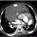
FIGURE 1.14 Occipital cephalocele in a girl at 30 weeks gestational age and 3-months of age. A: Sagittal HASTE fetal MRI obtained at 30 weeks demonstrates a suboccipital cephalocele (arrow) with question of a tongue of cerebellar parenchyma extending to the defect. B and C: Sagittal (B) and axial (C) FIESTA postnatal MR images demonstrate herniation of the left cerebellar hemisphere through the osseous/dural defect (arrow).

Parietal cephaloceles are the second most common site for cephaloceles in most populations.116,117,118 A type of rudimentary cephalocele known as an atretic cephalocele is particularly common in this location. These atretic cephaloceles communicate with the scalp through a thin fibrous connection and tiny skull defect. Classically, they are associated with upward deviation of the tentorium, a persistent falcine vein with underdeveloped straight sinus, a divided superior sagittal sinus, and faintly enhancing scalp mass consisting of rudimentary meningeal tissue (Fig. 1.18).122,123,124 When some of the classic imaging findings are absent and a skull defect is not appreciated, these atretic cephaloceles may be mistaken for a dermoid cyst or other soft tissue mass of the scalp.124

Basal cephaloceles involve the skull base and are further subdivided by location125,126: transsphenoidal, transethmoidal, sphenoethmoidal, transtemporal, spheno-orbital, and sphenomaxillary. Cephaloceles in this location typically present because of CSF leakage or recurrent infection (Figs. 1.17 and 1.19).126 Basal and temporal cephaloceles are the rarest cephaloceles encountered, constituting only 5% to 10% of all cephaloceles in most case series.118,127


FIGURE 1.15 Large interfrontal cephalocele and Tessier facial cleft in a 16-month-old girl. A and B: Axial (A) and sagittal reformatted (B) CT images demonstrate a large interfrontal cephalocele. The sagittal reformatted CT image demonstrates that the axis of herniation extends above the frontonasal suture (arrowhead). C and D: Frontal (C) and left lateral (D) perspective surface renderings of the CT data demonstrate the large frontal bone defect, the presence of the left paramedian Tessier facial cleft (black arrow), and some residual bone at the frontonasal suture (white arrow).

Only gold members can continue reading. Log In or Register to continue

Stay updated, free articles. Join our Telegram channel

Oct 13, 2018 | Posted by in PEDIATRIC IMAGING | Comments Off on Skull

Full access? Get Clinical Tree

Get Clinical Tree app for offline access