Heart


Heart


Lorna P. Browne

Edward Y. Lee

Oleksandr Kondrachuk

Marielle V. Fortier

Zhu Ming

Cynthia K. Rigsby


INTRODUCTION

In recent years, congenital and acquired forms of cardiac disease have been encountered with increasing frequency in the pediatric population. This is because more children with congenital heart disease survive longer mainly due to the substantial advances in surgical techniques and management. Radiologic imaging remains at the forefront of the diagnostic evaluation in children with clinically suspected or known congenital heart disease. However, the inherent complexity of these conditions continues to be a challenge for many radiologists.

The objective of this chapter is to enhance the understanding of congenital and acquired forms of cardiac disease occurring in the pediatric population by using a stepwise approach to cardiac anatomy and diagnosis that can be applied by the novice resident or expert radiologist. First, the various imaging techniques currently available for evaluating pediatric cardiac disease are discussed. The normal anatomy of the heart including the systemic and pulmonary arterial and venous vessels is reviewed. The beneficial use of the segmental approach to diagnosis is presented. Finally, a variety of the commonly encountered pediatric cardiac diseases focusing on pathophysiology, clinical features, imaging assessment, and treatment options are discussed.


IMAGING TECHNIQUES

Several imaging modalities are currently utilized in the evaluation of infants and children with suspected congenital or acquired cardiac disease, including radiography, echocardiography, computed tomography (CT), and magnetic resonance imaging (MRI), each of which has its own distinct advantages and disadvantages.


Radiography

Chest radiography is a useful initial evaluation in the infant or child with suspected cardiac disease, but its role in diagnosis has been supplanted by other imaging modalities. Still, many congenital cardiac conditions, such as tetralogy of Fallot (TOF), transposition of great arteries (TGA), supracardiac total anomalous pulmonary venous return (TAPVR), and Ebstein anomaly, have classical radiographic appearances.1 Currently, chest radiography is frequently used to screen pediatric patients with suspected underlying cardiac disease for assessment of cardiac size and evaluation of pulmonary vasculature. In addition, it is also used in pediatric patients with known cardiac disease in order to evaluate the pulmonary circulation for evidence of pulmonary venous congestion and response to intervention.


Echocardiography

Echocardiography (echo) is unrivaled in its ability to evaluate the intracardiac structures with excellent temporal and spatial resolution. It is commonly the first test performed in a neonate with suspected cardiac disease, usually before chest radiography. Limitations of echo include poor echo windows in older children or those with skeletal malformations and its inability to adequately visualize the extracardiac arterial and venous vasculature.2 In general, echo falls more within the purview of a pediatric cardiologist than a pediatric radiologist. Therefore, for the most part, this chapter is dedicated to the other (nonecho) imaging modalities that are currently used for evaluation of congenital and acquired forms of cardiac disease in the pediatric population.


Computed Tomography

Previously, pediatric cardiac computed tomography angiography (CTA) was associated with relatively long acquisition times and high radiation doses, limiting its utility in pediatric cardiac imaging. However, the advent of prospective ECG gating, ultrafast gantry rotation times, dual-source technology/volume acquisition, variable pitch (higher pitch with fast heart rate), and radiation dose modulation has enabled successful pediatric cardiac CTA with low radiation doses (˜1 to 3 mSv or less), frequently not requiring sedation or breath-holding.2 Although high heart rates (>100 bpm) are typical in infants and young children, beta blockade and vasodilation agents are not used routinely. Common applications of pediatric cardiac CTA include evaluation for anomalous coronary arteries, anomalous pulmonary veins, pulmonary artery stenosis/atresia, major arteriopulmonary collateral arteries (MAPCAs), and aortic root dilatation/dissection.2


Magnetic Resonance Imaging

The cardiac MRI evaluation of pediatric congenital heart anomalies can be subdivided into an assessment of cardiovascular morphology, quantification of ventricular function, and quantification of flow. Each of these assessments involves dedicated MRI sequences optimized for their individual role.3

Evaluation of cardiovascular morphology is performed using static spin-echo “black blood” sequences (T1/T2) or cine gradient-echo “bright blood” sequences (2D steady-state free precession [SSFP]). Spin-echo black blood techniques allow a static overview of the extracardiac thoracic vasculature. Cine gradient-echo imaging allows a dynamic assessment of the thoracic vessels with multiple frames acquired throughout the cardiac cycle. This provides a more accurate depiction of stenosis/aneurysms in vessels whose diameters are changing throughout the cardiac cycle.3,4 Coronary artery evaluation involves a specific sequence (3D SSFP), which yields high-resolution images only during a short period of each cardiac cycle when the heart is relatively motionless (usually end diastole for lower heart rates and end systole for higher heart rates). As this is a long sequence, typically taking 5 minutes or longer, respiratory motion is minimized by using a respiratory navigator during image acquisition. This technique is timed to have imaging occur during a small length of diaphragmatic excursion. Finally, contrast-enhanced 3D magnetic resonance angiography (MRA) also provides excellent morphologic assessment of the thoracic and abdominal vasculature with time-resolved techniques. Such techniques enable a fast acquisition that can be used to isolate the pulmonary and systemic arterial phases of contrast enhancement.5


FIGURE 9.1 Standard cardiac magnetic resonance imaging planes. Bright blood magnetic resonance images in (A) two-chamber, (B) four-chamber, and (C) short-axis geometries. LA, left atrium; LV, left ventricle; RA, right atrium; RV, right ventricle.

Evaluation of ventricular function is performed with cine 2D SSFP sequences that are optimized to provide excellent myocardial blood pool differentiation.3,4 Images are obtained in various planes that are similar to those acquired on echo and include a two-chamber plane (coronal oblique view demonstrating the right/left atrium and accompanying ventricle), a four-chamber plane (transverse oblique covering both atria and ventricles), and a short-axis plane (sagittal oblique across the right ventricle [RV] and left ventricle [LV]) (Fig. 9.1). Additional views, such as left and right outflow tract planes as well as aortic root planes (Fig. 9.2), may also be acquired depending on the pathology being evaluated.

Ventricular volume is calculated by summing the individual volumes of the ventricles on each of the short-axis or four-chamber slices acquired in end diastole and end systole, using specialized postprocessing software. Wall motion abnormalities are demonstrated by visualization of the images in a dynamic mode throughout the cardiac cycle. Immediately following the intravenous administration of gadolinium, first-pass perfusion sequences may show areas of delayed or absent myocardial perfusion related to ischemia or infarction, respectively.4,5 Administration of a pharmacologic stress agent such as adenosine or dobutamine (stress perfusion) can be utilized to elicit decreased first-pass perfusion that is not
visible under resting conditions. In general, adenosine is preferred because of its ease of administration, fast action, and short half-life. However, unlike in adults, stress perfusion in pediatrics is usually reserved for those patients clinically felt to be high risk for myocardial ischemia, such as those with coronary artery involvement in Kawasaki disease or in heart transplant patients with suspected coronary artery vasculopathy. Approximately 10 minutes after administration of gadolinium, myocardial viability sequences can be performed to demonstrate retained gadolinium in regions of myocardial fibrosis (Fig. 9.3).


FIGURE 9.2 Outflow tract cardiac magnetic resonance imaging planes. Bright blood magnetic resonance images in (A) left ventricular outflow tract/three-chamber view (B) right ventricular outflow tract. AA, ascending aorta; LA, left atrium; LV, left ventricle; MPA, main pulmonary artery; RV, right ventricle.

Flow evaluation is performed with cine phase-contrast sequences (Fig. 9.4). Flow sequences are used to assess stroke volumes and regurgitant fractions across the aortic and pulmonary valves. In addition, flow sequences can be used to measure the volume of systemic to pulmonary shunting, known as the Qp:Qs ratio where Qp is the volume of pulmonary blood flow and Qs is the volume of systemic blood flow.4,5 The Qp:Qs ratio is an important quantification in the evaluation of septal defects and other left-to-right shunts. Pressure gradients across stenoses may be derived using the modified Bernoulli equation (4 × V2 = ΔP, where ΔP is the pressure gradient in mm Hg and V is the measured peak velocity by cine phase contrast in m/s).

The entire cardiac MRI examination, including a morphologic assessment, ventricular function, and flow analysis, is expected to take between 60 and 90 minutes; therefore,
frequently, general anesthesia or conscious sedation is necessary in the pediatric population.


FIGURE 9.3 Delayed cardiac wall enhancement. Short-axis plane phase-sensitive inversion recovery magnetic resonance image shows an area of subendocardial enhancement (arrow) in the anterolateral wall of the left ventricle.


FIGURE 9.4 Flow quantification using phase-contrast angiography. Phase contrast across the main pulmonary artery (MPA) demonstrates an approximately 40% pulmonary regurgitant fraction (green circle on A and arrow on B).


NORMAL ANATOMY

A clear understanding of normal cardiac anatomy is fundamental to the imaging interpretation of congenital heart anomalies and abnormalities.

The normal pediatric heart occupies ˜60% of the cardiothoracic diameter on chest radiography in the neonatal period and early infancy, decreasing to about 50% in later childhood life and adolescence. When evaluating both normal and complex cardiovascular anatomy on cross-sectional imaging techniques such as CT or MRI, a segmental approach, which can enhance interpretation, is recommended.6 This approach broadly encompasses a series of steps that assess the positions of the cardiac chambers and great vessels and their relationships in a sequential fashion, as if the interpreter were a red blood cell traveling from the systemic veins into the heart and ultimately terminating in the systemic arteries.

The first step is the evaluation of visceral situs. This involves confirmation of rightward location of the liver and inferior vena cava (IVC) and leftward location of the spleen and stomach. The cardiac position (referred to as cardiac situs) is determined by the position of the cardiac apex, which is normally inferior and leftward and termed levocardia. Mesocardia refers to a heart in a more midline location. Dextrocardia indicates that the heart is located in the right chest with the apex pointing rightward. In dextroversion, the heart is shifted rightward, either through mass effect or pulmonary volume loss, but the cardiac apex still points to the left.

The next step involves evaluation of the systemic venous anatomy, including confirmation of a single right-sided superior vena cava (SVC), normal position of the right and left innominate veins, and the presence of a normal suprarenal IVC. Commonly encountered systemic venous anomalies include bilateral SVCs (usually without a left innominate vein) and an interrupted IVC with azygos continuation.

The atrial situs evaluation comes next. It is the morphology of the atria and their venous inflow, rather than their location in the chest, which determines whether the atria are termed right or left. The atrium that receives the suprahepatic IVC and has a broad-based atrial appendage is considered to be the right atrium. The SVC is more variable in its drainage than the suprahepatic IVC. Therefore, it is not used in determination of atrial morphology. The atrium that has a long and narrow atrial appendage is termed the left atrium and normally should receive the four pulmonary veins.

The determination of ventricular morphology can establish what type of ventricular looping took place during embryonic cardiac development and also the type of atrioventricular valve (AVV) supplies the ventricles. The RV has a number of prominent trabeculations along the septum and ventricular free wall. One of these trabeculations is particularly prominent and is known as the moderator band (Fig. 9.5). The moderator band runs from the ventricular free wall to the interventricular septum and is relatively closely located to the cardiac apex. The moderator band can be easily identified on an axial CT view of the chest and a four-chamber view
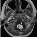
on cardiac MRI and is a reliable determinant of ventricular morphology. The tricuspid valve follows the RV. Typically, the tricuspid valve is located very slightly closer to the cardiac apex than the mitral valve. The tricuspid valve has three leaflets, including the septal leaflet (attached to the interventricular septum) and anterior and posterior leaflets. The LV is supplied by the mitral valve and typically has two papillary muscles along its free wall (anterolateral and posteromedial). However, these papillary muscles are more variable in appearance than moderator band in terms of correctly identifying ventricular morphology. If the RV is located anteriorly and rightward relative to the LV, then normal ventricular “D” looping is deemed to have occurred. If the ventricle containing the moderator band is located posteriorly and leftward, then ventricular inversion or “L” looping has occurred. This is discussed in more detail later in the chapter in TGA.


FIGURE 9.5 Moderator band. Bright blood magnetic resonance image in four-chamber plane shows the moderator band (arrow) in the right ventricle extending from the free wall to the ventricular septum. LV, left ventricle; RV, right ventricle.

Evaluation of the great arteries starts with evaluation of the conus (infundibulum). The conus is the muscular sleeve that is normally located under the pulmonary valve around the infundibulum of the heart. It forms the superior portion of the interventricular septum. In addition, it separates the pulmonary valve from the AV and the aortic valves. Its presence, absence, or variation in appearance forms a central role in the development of complex malformations such as TOF, interrupted arch, TGA, and double outlet right ventricle (DORV).

The next step is the assessment of great vessel arrangement and ventriculoarterial connections. Normally, the LV is attached to the aorta, and the RV is attached to the main pulmonary artery (MPA). If this normal arrangement is present, the ventriculoarterial connection is described as concordant. If the LV is connected to the pulmonary artery and the RV is connected to the aorta, the arrangement is described as discordant. The normal relationship of the great arteries results in the aortic valve annulus located rightward and posterior to the pulmonary valve annulus (termed situs solitus of the great vessels). Situs inversus of the great vessels is the term used when the aortic annulus is located leftward and posterior of the pulmonary valve yet normal ventriculoarterial concordance is maintained (i.e., LV → Aorta and RV → MPA). In discordant ventriculoarterial connections (i.e., LV → MPA and RV → Aorta), the great vessels are described as being transposed. In this case, the aortic valve annulus may be either rightward and anterior to the pulmonary valve (D-transposed) or leftward and anterior to the pulmonary valve (L-transposed).

Once the cardiac segmental anatomy is established, the interatrial and interventricular septa are assessed for defects, and the great vessels are interrogated for stenoses or other abnormalities.

Approaching the cardiac anatomy using the above segmental approach greatly assists in correctly identifying and classifying cardiac malformations.6


Atria

The atria are the cardiac chambers that receive the systemic and pulmonary venous drainage. The right atrium forms the right heart border of the cardiac silhouette on frontal chest radiographs. It receives systemic venous blood from the two venae cavae and from the coronary veins via the coronary sinus. Then the right atrium expels blood through the tricuspid valve into the RV. The interior of the right atrium has a smooth posterior wall and a muscular anterior wall that is separated by a crescent-shaped muscular ridge, the crista terminalis. The inferior vena caval orifice is guarded by the Eustachian valve and the coronary sinus by the Thebesian valve. Both the inferior vena caval orifice and the coronary sinus orifice are located along the inferior border of the atrium. The superior vena caval orifice is valveless, but the pacemaker of the heart, the sinoatrial (SA) node, is located just posterior to the superior vena caval orifice.

The left atrium forms the posterior border of the heart on lateral chest radiographs. The left atrium is located just below the carina and left main bronchus. Such location of the left atrium with respect to the carina and left main bronchus explains why the carina appears splayed and left main bronchus appears elevated on frontal chest radiographs in cases of severe left atrial enlargement. The left atrium typically receives at least four pulmonary veins and expels blood through the mitral valve into the LV. There is a normal ridge of tissue known as the Coumadin ridge, which separates the left upper pulmonary vein from the left atrium that is confluent with the wall of the left atrial appendage. This should not be confused with cor triatriatum.7


Atrial Septum

The interatrial septum not only separates the left and right atria but also has a small portion that separates the right atrium from the LV, known as the AV septum. The importance
of the AV portion is that it represents one of the boundaries of the triangle of Koch that is the anatomic landmark for the AV node. In addition to the SA node, the AV node forms part of the conduction system of the heart. Along the midportion of the interatrial septum lies a small depression known as the fossa ovalis/foramen ovale that represents the remnant of the ostium secundum and should close shortly after birth.7


Ventricles

The RV forms most of the inferior border of the cardiac silhouette on frontal chest radiographs. Normally, the RV is anterior and rightward with respect to the LV. The RV can be divided into inlet, trabecular, and outlet portions. The inlet portion is derived in association with the tricuspid valve and contains the chordal attachments. The trabecular portion contains prominent muscle bundles that traverse the chamber from the free wall to the interventricular septum and include the moderator band. Other important named muscle bundles include the parietal band and the septal band. The parietal band separates the tricuspid and pulmonary valves. The septal band, which is Y-shaped, merges with apical trabeculations, gives rise to the moderator band (Fig. 9.5), and ends in the tricuspid papillary muscle. These three muscle bands form a circular ring known as the crista supraventricularis, which separates the trabecular portion of the ventricle from the outlet portion. The outlet portion of the RV is contained within a collar of muscle known as the conus arteriosus (“conus,” or infundibulum), and it separates the pulmonary and tricuspid valves.

The LV forms the left border of the heart on frontal chest radiographs. The muscle bundles of the septal and free walls of the LV are arranged in a spiral fashion and also crisscross one another. As a consequence, systole results in twisting contractions, which “wring” the blood out of the LV. The LV is also divided into inlet, trabecular, and outlet portions. The inlet portion of the LV is short and contains the mitral chordal attachments. The trabecular portion contains the anterolateral and posteromedial papillary muscles and fine apical trabeculations. The outlet portion is quite long and does not have a conus infundibulum muscular ring (as is seen in the right ventricular outlet), so the aortic and mitral valves normally are in fibrous continuity.


FIGURE 9.6 Normal coronary arteries. A: Axial maximum intensity projection (MIP) CT image demonstrates normal origin of the right coronary artery (RCA) and left coronary artery (LCA) from the right (R) and left (L) sinuses of Valsalva. (N, noncoronary sinus.) B: Three-dimensional volume-rendered CT image shows normal courses of the RCA, LCA, left anterior descending coronary artery (LAD), and left circumflex coronary artery (CIRC).

Division of the LV into four segments is used in imaging when assessing myocardial function and evaluating for perfusion defects. In this segmentation scheme, the LV is considered to be a circular structure, and the four segments are septal (interventricular septum), anterior (superior wall), inferior (inferior wall), and lateral (mid free wall). The locations where these quadrants overlap are termed anteroseptal, anterolateral, inferoseptal, and inferolateral.7


Interventricular Septum

The ventricular septum is divided in a similar way to the ventricular chambers. The inlet septum is bordered by tricuspid valve attachments, the outlet septum lies superior to the crista supraventricularis, and the trabecular septum is located between the inlet and outlet portions. Such division is useful when trying to localize ventricular septal defects (VSDs). One additional portion of the outlet septum is termed the membranous septum, which is a very small portion of the septum that lies between the pulmonary valve annulus and
inferior aspect of the tricuspid valve annulus. The AV bundle (of HIS) runs through the membranous septum and is the only normal route for connection between the AV node and the ventricular myocardial conduction system.7


Coronary Arteries

The right and left main coronary arteries (RCA and LCA) originate from the center of the right and left aortic sinuses of Valsalva, which are located on either side of the aortic root, facing the pulmonary valve8 (Fig. 9.6). The third aortic sinus, known as the noncoronary sinus, is located posterior to the other two aortic sinuses. The right and left coronary arteries arise from their respective aortic sinuses about halfway between the aortic valve annulus and the sinotubular junction. The right coronary artery (RCA) travels in the right AV groove and supplies the right ventricular free wall via a conal branch and multiple marginal branches. The left coronary artery rapidly divides into left anterior descending (LAD) and circumflex branches. The LAD artery travels in the anterior interventricular groove. It usually supplies the myocardium of the left ventricular free wall via diagonal branches and the anterior interventricular septum via septal branches. The circumflex artery travels within the left AV groove. It terminates in obtuse marginal branches that supply the lateral wall of the LV and part of the anterolateral papillary muscle.8

The artery that gives rise to the posterior descending artery (PDA), which supplies the inferior interventricular septum, inferior left ventricular free wall, and the AV node, is considered to be the dominant coronary artery. In ˜70% of individuals, the RCA gives rise to the PDA (right coronary dominant); in 10%, the circumflex artery gives rise to branches to the posterior right ventricular surface (left circumflex [LCX] dominant); and in the remaining 20%, the PDA is supplied by branches from both right coronary and LCX arteries (codominant).8


CONGENITAL CARDIAC MALFORMATIONS


Septal Defects


Atrial Septal Defects

Atrial septal defect (ASD) arises from failure of closure of portions of the interatrial septum (ostium primum and ostium secundum) or maldevelopment of the portion of the atrium that receives the vena cava (sinus venosus defects). There are three major types of ASDs: ostium primum, ostium secundum, and sinus venous defects9 (Fig. 9.7).

The atrial septum is formed from the septum primum. This starts in the roof of the primitive common atrium and grows toward the endocardial cushions, located between the primitive atria and ventricles. The gap between the septum primum and the endocardial cushion is known as an ostium primum. The gap gradually decreases as the septum primum and the endocardial cushions fuse to form the interatrial septum and atrioventricular valves, respectively. If the septum primum and endocardial cushions fail to fuse completely, an ostium primum ASD results (10% to 15% of ASDs), which is an ASD that is located close to the AV (mitral and tricuspid) valves. It is usually associated with maldevelopment of the atrioventricular valves (AVVs).9


FIGURE 9.7 Diagram of the location of the three types of atrial septal defects. ASD 1: primum atrial septal defects. ASD 2: secondum atrial septal defects. ASD 3: sinus venosus superior/inferior atrial septal defects.

The ostium secundum results from small perforations, which form in the septum primum and which gradually coalesce. The ostium secundum is usually partially covered by a valve of tissue formed by a fold in the septum primum. This fold (septum secundum) usually fuses completely with the rest of the interatrial septum shortly after birth closing the ostium secundum. Persistence is known as a patent foramen ovale or ostium secundum ASD comprising of 80% of all ASDs (Fig. 9.8).9

The sinus venosus is the posterior portion of the right atrium formed by the incorporation of primitive cardinal veins into the primitive atrium.10 Sinus venosus defects (5% to
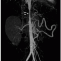
10% of all ASDs) occur as a result of either malposition of the interatrial septum or malposition of the SVC/IVC resulting in a vena cava that overrides the atrial septum. Therefore, a sinus venous defect is actually a defect above or below the interatrial septum and not truly within the septum like ostium primum and secundum defects. A superior sinus venous defect where the SVC meets the interatrial septum is much more common than the inferior sinus venosus type. The resulting gap between the interatrial septum and the vena cava causes pulmonary venous blood in the left atrium to be drawn into the lower pressure right atrium. In addition, at least 85% patients with superior type sinus venosus defects have concomitant anomalous partial anomalous pulmonary venous drainage from the right upper lobe into the SVC (Fig. 9.9).


FIGURE 9.8 Ostium secundum atrial septal defect. Bright blood magnetic resonance image in four-chamber plane shows ostium secundum atrial septal defect (arrow).

All ASDs result in a left-to-right shunt, which may lead to complications. A small ostium secundum defect may close spontaneously in the first 2 years of life. The remaining ASDs or ostium secundum ASDs in patients older than 2 years of age are unlikely to close. An untreated large ASD leads to increased pulmonary blood flow, right atrial and right ventricular enlargement, and pulmonary hypertension.9

On chest radiographs, cardiomegaly and pulmonary vascular congestion with an enlarged central pulmonary vessels and variable amounts of pulmonary edema depending on the size of the shunt are seen. Currently, cardiac gated CTA is occasionally used to assess ostium secundum ASDs for possible percutaneous septal device closure. A 5 mm rim of tissue separating the ASD from each of the following structures: aortic annulus, SVC orifice, IVC orifice, right upper pulmonary venous orifice and the atrioventricular valve is needed for successful device placement. Cardiac MRI can evaluate the presence and location of the ASD, which is close to the tricuspid and mitral valves in ostium primum defects, midinteratrial septum in ostium secundum defects, and posterior aspect of cavoatrial junction in sinus venous defects. It also provides information regarding the size of the rim of septal tissue that surrounds an ostium secundum ASD, which helps determine candidacy for percutaneous closure. In addition to the assessment of right ventricular function, cardiac MRI can also quantify the shunt volume or Qp:Qs (ratio of volume of blood in the pulmonary circulation: volume of blood in the systemic circulation) by comparing stroke volumes in the RV and MPA to stroke volumes in the LV and aorta. Furthermore, cardiac MRI provides excellent evaluation of the pulmonary veins given the association of anomalous pulmonary venous drainage in sinus venosus type defects.


FIGURE 9.9 Sinus venous defect. A: Axial bright blood magnetic resonance (MR) image demonstrates a superior sinus venous defect (asterisk) between the posterior wall of superior vena cava (SVC) and anterior left atrium (LA). B: Three-dimensional volume-rendered MR image shows partial anomalous pulmonary venous return (arrow) from right upper lobe to SVC.

Ostium secundum defect closure can be achieved either surgically using a pericardial/Gore-Tex patch or percutaneously using a septal closure device in select cases.11 Surgical ostium primum defect closure involves placing a patch. The type of surgical repair undertaken for repairing sinus venosus defects is dependent upon the location of the drainage of the anomalous pulmonary veins. The Warden procedure is generally performed when the anomalous pulmonary veins return to the SVC. This procedure involves oversewing the SVC above the anomalous pulmonary venous connection and anastomosing the proximal end of the SVC to the right atrial appendage.10 The anomalous pulmonary vein(s) and caudal end of the SVC are baffled to the left atrium through the sinus venosus defect thus closing the defect. A single patch closure of the sinus venosus defect is performed if the anomalous pulmonary veins drain to the right atrium or SVC/right atrial junction.12


Atrioventricular Septal Defects

The two endocardial cushions are located between the primitive atria and ventricles. They normally fuse to form the mitral and tricuspid valves and the crux of the heart, at the junction of the interatrial and interventricular septa.13 Atrioventricular
septal defects (AVSDs) are malformations caused by failure of fusion of the endocardial cushions that result in a deficient AV septum with a common atrioventricular valve (AVV) rather than separate mitral and tricuspid valve openings.14 This common AVV often has five to six leaflets and a complex arrangement of chordal attachments. Normally, the aortic valve is situated between the tricuspid and mitral valves. However, when there is common AVV, the aortic valve becomes displaced superiorly, resulting in an elongated and potentially narrowed left ventricular outflow tract.

If the AVV is positioned centrally over the ventricles, it is termed a balanced AV septal defect and results in a large left-to-right shunt. An unbalanced AV septal defect results when the common AVV is located more over one ventricle than the other. This affects ventricular development and a single ventricle physiology usually results. For instance, if the AVV is situated more over the RV, then LV becomes underdeveloped and a hypoplastic left heart physiology results. Such location of the AVV may be exacerbated by coexistent left ventricular outflow tract stenosis resulting from the displaced aortic valve. This is discussed more fully in the single ventricle portion of the chapter.

AVSDs result in significant left-to-right shunting. Therefore, clinical presentation is in the early neonatal period with respiratory distress, murmur, and congestive cardiac failure. AVSDs are commonly associated with Down syndrome and anomalies of atrial and visceral situs.15

Chest radiography demonstrates global cardiomegaly (Fig. 9.10), sometimes with a horizontal superior right atrial border, severe pulmonary vascular engorgement, and pulmonary edema.1 CT usually has no role in the evaluation of AVSDs. Cardiac MRI is usually reserved for complex cases not completely assessed on echo. In these cases, cardiac MRI can quantify shunt volume and AVV regurgitation, depict complex intracardiac anatomy, and determine ventricular size and function, which may facilitate presurgical planning (Fig. 9.11).


FIGURE 9.10 Balanced atrioventricular septal defect. Frontal chest radiograph shows increased pulmonary vascularity and global cardiomegaly.


FIGURE 9.11 Balanced atrioventricular septal defect. Bright blood magnetic resonance image in four-chamber plane demonstrates large atrioventricular septal defect (asterisk). LA, left atrium; LV, left ventricle; RA, right atrium; RV, right ventricle.

Surgical correction of balanced AVSDs can be achieved by closure of the ostium primum ASD and inlet VSD using one or two patches, which are then used to recreate separated atrioventricular valves.15,16 Surgical correction of unbalanced defects depends on the type of physiology and is discussed in the single ventricle portion of the chapter.


Ventricular Septal Defects

VSDs are holes in the interventricular septum. They can occur as an isolated defect or be part of more complex congenital heart anomalies such as conotruncal anomalies (TOF, TGA, DORV) or AVSDs. VSDs are the second most commonly encountered congenital heart defects after bicuspid aortic valves.17

The interventricular septum first appears as a muscular ridge close to the cardiac apex at the end of the 4th week. Then, it grows cephalad as the RV and LV enlarge, eventually fusing with bulbus cordis (primitive ventricular outflow tract). The inlet portion of the ventricular septum is thought to arise from the endocardial cushions. The membranous portion of the ventricular septum forms last and results from the formation of fibrous valvar leaflets arising from the endocardial cushions. The union of the endocardial cushions with the muscular interventricular septum is still not completely understood with differing hypotheses. However, final fusion between the muscular septum, membranous septum, and inlet septum occurs at approximately the 8th gestational week.

There are four types of VSDs that reflect the embryologic development of the interventricular septum: inlet, muscular, perimembranous, and subarterial17 (Fig. 9.12). The inlet type of VSD extends from the fibrous annulus of the AVV and is often associated with an endocardial cushion defect. Muscular defects may appear anywhere throughout the muscular portion of the ventricular septum. Small muscular VSDs
usually close spontaneously, and larger VSDs generally require surgical intervention. Occasionally, the muscular part of the septum can have innumerable VSDs, resulting in the so-called Swiss cheese interventricular septum. Perimembranous VSDs are the most common type to require surgical intervention and are characterized by a defect in the membranous portion of the ventricular septum. The subarterial or malalignment type of VSD appears just below the aortic valve cusps. Perimembranous and subarterial VSDs are often referred to as outlet VSDs and are associated with conotruncal anomalies such as TOF and TGA.17


FIGURE 9.12 Diagram of the location of the four types of ventricular septal defects (VSDs) including subpulmonary VSD (subarterial), membranous VSD (perimembranous), atrioventricular canal defect (inlet type), and muscular VSD (muscular).

Clinical presentation depends on the size of the defect and whether other cardiac anomalies are present. Small isolated VSDs often require no treatment and usually spontaneously close during childhood, whereas larger defects may manifest with failure to thrive and signs of congestive heart failure. Chronic left-to-right shunting may result in pulmonary hypertension leading to reversal of shunting known as Eisenmenger syndrome.18

Chest radiography appearances of VSDs depend of the size of the VSD and vary from normal cardiac size to severe cardiomegaly with normal pulmonary vascularity to severe pulmonary vascular engorgement and pulmonary edema (Fig. 9.13). CT has no role in the evaluation of VSDs although they are sometimes found incidentally (Fig. 9.14). Cardiac MRI is used usually reserved for evaluation of complex cases, which are not completely assessed on echo (Fig. 9.15). In these cases, cardiac MRI can quantify shunt volume, depict associated intracardiac or valvar anomalies, and determine ventricular size and function, which may facilitate presurgical planning.


FIGURE 9.13 Ventricular septal defect. Frontal chest radiograph shows increased pulmonary vascularity and cardiac enlargement.

Surgical closure is favored for treatment of VSDs that are unlikely to close spontaneously or are associated with a significant left-to-right shunt. This is usually performed using a patch of synthetic material.18 Percutaneous device closure of these defects is rarely performed because of the reported incidence of both early- and late-onset complete heart block after device closure of perimembranous VSDs, which occurs presumably secondary to device trauma to the adjacent AV node. Occasionally, percutaneous closure of muscular VSDs is performed using a septal occluder device.19


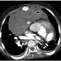
FIGURE 9.14 Ventricular septal defect. Axial enhanced CT image shows a large muscular VSD (arrow), partially covered by right ventricular muscle bundles. LV, left ventricle; RV, right ventricle.


FIGURE 9.15 Perimembranous ventricular septal defect. Black blood magnetic resonance image shows a perimembranous ventricular septal defect (arrow) with muscular extension in a 2-month-old girl with double outlet right ventricle.


Atrioventricular Valve Stenosis and Regurgitation


Mitral Stenosis

Congenital mitral stenosis is very rare and usually occurs in association with other left heart lesions including hypoplastic left heart syndrome (HLHS) and aortic stenosis/atresia. Asymmetric congenital mitral stenosis is termed parachute mitral valve, which may be associated with a single papillary muscle with fused chordal attachments, giving the mitral valve an unbalanced appearance.7 The most common cause of acquired mitral stenosis in young patients is rheumatic heart disease (Fig. 9.16). Patients with mild stenosis are usually asymptomatic, whereas patients with more severe disease develop pulmonary venous congestion.

Chest radiography may be normal or demonstrate pulmonary vascular congestion and interstitial edema depending on the degree of underlying obstruction. Cardiac MRI and CTA currently do not usually have a role in evaluating mitral stenosis.


FIGURE 9.16 Mitral stenosis. Axial bright blood magnetic resonance image shows turbulent jets arising from the tips of stenosed mitral valve leaflets (arrows). LV, left ventricle.

Surgical repair of congenital mitral stenosis involves valvuloplasty and often later valve replacement surgery.


Mitral Regurgitation

Congenital mitral regurgitation is very rare. It is usually seen in association with a cleft in the mitral valve, connective tissue disorders such as Marfan syndrome, or secondary to ventricular dysfunction, trauma, or myocardial infarction.7 Mitral valve prolapse is thought to be related to an inherent abnormality in the myxomatous matrix/collagenous structure of the mitral valve.20 Clinically, symptoms depend on severity of disease. Infants and children with severe disease usually demonstrate symptoms of pulmonary vascular congestion at <3 years of age.

Chest radiography may reveal elevation of the left main bronchus, splaying of the carina, and an enlarged posterior cardiac shadow on the lateral projection related to left atrial enlargement. Although echo is the primary diagnostic test for evaluating mitral regurgitation, cardiac MRI is frequently used in the overall cardiovascular assessment in patients with connective tissue disorders such as Marfan syndrome. Mitral regurgitation can be seen as a regurgitant jet arising from the valve leaflets during ventricular systole, usually resulting in left atrial dilatation (Fig. 9.17). Mitral valve prolapse is best appreciated on the left ventricular outflow tract/three-chamber view. This view shows the anterior mitral valve leaflet prolapsing into the atrium during systole with an accompanying regurgitant jet into the left atrium (Fig. 9.18).


FIGURE 9.17 Mitral regurgitation. Two-chamber plane magnitude phase-contrast magnetic resonance image demonstrates mitral valve regurgitation jet (arrow) into an enlarged left atrium (LA). LV, left ventricle.


FIGURE 9.18 Mitral valve prolapse. Bright blood magnetic resonance image in left ventricular outflow tract plane shows mitral valve leaflets (asterisks) prolapsing into the left atrium (LA) with a prominent regurgitant jet (arrow). AA, ascending aorta; LV, left ventricle.

A cleft in the mitral valve can usually be surgically repaired either primarily or with a patch. Occasionally, annular plication is used in severe mitral valve prolapse with redundant leaflets. Mitral valve replacement is occasionally necessary in pediatric patients with severe mitral regurgitation or those who have failed more conservative surgical measures.


Tricuspid Stenosis

Congenital tricuspid stenosis is usually associated with other anomalies of the right heart including right ventricular hypoplasia and right ventricular outflow tract (RVOT) obstruction/pulmonary atresia (Fig. 9.19). Isolated tricuspid stenosis is extremely rare. Tricuspid stenosis is similar to tricuspid atresia clinically, on imaging, and surgically.7


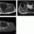
FIGURE 9.19 Tricuspid stenosis. Axial enhanced CT image shows thickened tricuspid valve with narrowed valve annulus (asterisk) and hypoplastic right ventricle (RV). Also noted is an extracardiac Fontan (F) shunt.


Tricuspid Regurgitation

Primary tricuspid valve dysplasia resulting in tricuspid regurgitation is most common in the setting of Ebstein anomaly and TGA. However, secondary tricuspid regurgitation may be seen in any congenital or acquired cardiac disease that leads to right ventricular volume overload, arising from secondary tricuspid annular distortion, and dilatation.7 Clinically, affected pediatric patients present with variable signs of right heart failure depending on the degree of underlying regurgitation.

Chest radiography may show right heart prominence related to right atrial enlargement. Cardiac MRI and CT are primarily used to identify the secondary causes of tricuspid regurgitation leading to right ventricular dysfunction (Fig. 9.20).


Ebstein Anomaly

Ebstein anomaly is a relatively rare and isolated anomaly of the tricuspid valve in which there is displacement of the septal and posterior tricuspid valve leaflets beyond the tricuspid annulus into the RV21,22 (Fig. 9.21). The portion of the RV between the tricuspid valve annulus and the displaced leaflets forms an “atrialized” portion of the right ventricular chamber (Fig. 9.22). The tricuspid valve is usually markedly dysplastic and regurgitant. In most cases, there is also an ASD or patent foramen ovale.21,22

The majority of affected pediatric patients present with cyanosis. Cyanosis is worst in the neonatal period where the functionally small RV may not be able to generate sufficient pressures to overcome the high pulmonary vascular resistance present after birth. After the pulmonary vascular resistance drops, typically around the 6th week, the cyanosis typically resolves only to reappear in the adolescent period, thought to be due to worsening tricuspid regurgitation.21

Chest radiography classically demonstrates a moderately to severely enlarged right heart with diminished pulmonary
vascularity1,22 (Fig. 9.23). Echo is used for initial diagnosis. However, MRI is playing an increasing role in the presurgical and postrepair assessment of right ventricular volumes, tricuspid valve morphology/regurgitation, and associated atrial or ventricular shunts.


FIGURE 9.20 Tricuspid regurgitation. Axial bright blood magnetic resonance image in four-chamber plane shows tricuspid regurgitant jet (arrow) and enlarged right atrium (RA). RV, right ventricle.


FIGURE 9.21 Diagram of the anatomy of Ebstein anomaly. The tricuspid valve is downwardly displaced and adherent to the interventricular septum. Part of the right ventricle is “atrialized,” being located superior to the tricuspid valve. LA, left atrium; LV, left ventricle; RA, right atrium; RV, right ventricle.

Surgical repair involves tricuspid valve plication, annuloplasty, or tricuspid valve replacement. In the neonatal stage, if cyanosis is severe, a palliative modified Blalock-Taussig (BT) shunt maybe required to supplement pulmonary arterial blood supply.22


Complex Univentricular Connections

Complex univentricular connections are congenital malformations that result in the atrioventricular valvar unit emptying into one ventricular chamber. These entities include HLHS, tricuspid atresia, double inlet LV, and unbalanced AVSDs. In these cases, the second ventricular chamber that is not supplied by an AVV is hypoplastic or atretic. The AVV unit may be composed of one common AVV, a normal-appearing mitral and tricuspid valve, or either a mitral or tricuspid valve. If present, the hypoplastic second ventricular chamber that is not supplied by an AVV is connected to the main ventricle by a VSD.


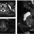
FIGURE 9.22 Ebstein anomaly. Bright blood magnetic resonance images in a four-chamber plane (A) and left ventricular outflow tract plane (B) show downward displacement of the attachment of the septal leaflet (straight arrow). The anterior leaflet (curved arrow) is large and “sail-like” resulting in an atrialized portion of the right ventricle (APRV).


FIGURE 9.23 Ebstein anomaly. Frontal chest radiograph demonstrates enlarged right cardiac margin.


Hypoplastic Left Heart Syndrome

HLHS is the most common single ventricle anomaly. HLHS has a series of congenitally small left-sided structures including a stenosed/atretic mitral valve, small LV, stenosed/atretic
aortic valve, small ascending aorta, and tubular hypoplasia of the aortic arch and aortic coarctation (Figs. 9.24 and 9.25).23 If there is severe aortic valve stenosis/atresia, then the ascending aorta and aortic arch may demonstrate retrograde filling from the ductus arteriosus, rather than anterograde flow through the aortic valve. In this situation, the coronary arteries also rely on retrograde flow leading to chronic myocardial hypoperfusion. Most of the oxygenated pulmonary venous blood crosses through an ASD into the pulmonary artery via the right heart and reaches the systemic arterial circulation through the ductus arteriosus.23,24 The descending aorta is usually normal caliber because of the presence of an enlarged ductus arteriosus. Clinical presentation is during the first few weeks of life coinciding with the closure of the ductus arteriosus. Manifestations vary depending on the degree of left-sided obstruction and include failure to thrive, hypoper-fused extremities, cardiac failure, and metabolic acidosis.7,23


FIGURE 9.24 Diagram of hypoplastic left heart associated with aortic atresia. The left ventricle (LV) is hypoplastic and hypertrophied. The ascending aorta (Ao) is very hypoplastic. LA, left atrium; LPA, left pulmonary artery; RA, right atrium; RPA, right pulmonary artery; RV, right ventricle.


FIGURE 9.25 Hypoplastic left heart syndrome. A: Axial enhanced CT image shows mitral valve annular stenosis (asterisk) and hypoplastic left ventricle (LV). RA, right atrium; RV, right ventricle. B: Sagittal reformatted enhanced CT image demonstrates tubular hypoplasia of ascending aorta (black arrow) and coarctation (white arrow) at aortic isthmus.

On chest radiography, the cardiac size can be normal or increased and there is reduced size of the aortic knuckle (Fig. 9.26). Additionally, there is usually pulmonary vascular congestion that is increased in the presence of a restrictive ASD. Echo is used to clearly delineate the intracardiac anatomy in the prerepair setting of HLHS. Once diagnosed, affected infants usually undergo immediate palliative surgery without additional imaging.

Cardiac MRI has a limited role in the preoperative assessment of HLHS. However, it can provide useful information in pediatric patients with less severe forms of left-sided obstruction and larger LVs who may benefit from a biventricular type of cardiac repair rather than a univentricular type of palliation.25 In these instances, cardiac MRI may be used to estimate the left ventricular end diastolic volume (LVEDV) and screen for the presence of endocardial fibroelastosis, which is used as a predictor of potential outcome. Predictors of a failed biventricular repair include a small LV (LVEDVs indexed to body surface area of <15 to 20 mL/m2), large VSD with right-to-left systolic shunting, severe mitral annular hypoplasia, and a dysplastic aortic valve.25 Other indications for CTA or MRA in pediatric patients with HLHS include aortic arch assessment, in order to assess for aortic coarctation and tubular arch hypoplasia.

Biventricular repair is reserved for a subset of children with the mildest forms of HLHS and least aortic and mitral valvar stenosis.25 Biventricular repair involves reconstruction of the ascending aorta with the MPA, a RV-PA conduit, and closure of the atrial and ventricular septal defects.

In the majority of patients with HLHS, the LV is too small to function as the systemic ventricle. Therefore, a staged single
ventricle palliation is performed,23,24 which results in the RV functioning as the systemic ventricle. This staged complex surgical procedure is performed in three stages, known as the Norwood I, the Glenn shunt (occasionally referred to as superior cavopulmonary anastomosis/Norwood II), and the Fontan shunt (occasionally referred to as Norwood III/total cavopulmonary anastomosis) procedure.23,24


FIGURE 9.26 Hypoplastic left heart syndrome. Frontal chest radiograph in a 1-day-old girl shows increased pulmonary vascularity and cardiomegaly. Aortic knuckle is not clearly visualized.

The first stage, known as the Norwood I procedure, is performed immediately after birth (Figs. 9.27 and 9.28; Table 9.1).

This procedure sacrifices the MPA in order to refashion an ascending aorta capable of sustaining the systemic arterial supply. The MPA is resected and separated from the right and left pulmonary arteries. The MPA is then used to augment the hypoplastic ascending aorta and proximal arch (the Damus-Kaye-Stansel maneuver). As the MPA has been resected, pulmonary arterial supply has to be reestablished. This can be accomplished using either a modified BT shunt from the right subclavian artery to the right pulmonary artery (RPA) (Fig. 9.28) or modified Sano shunt (RV) to confluence of branch pulmonary arteries (Fig. 9.29).23,24


FIGURE 9.27 Diagram of the Norwood stage 1 operation. The pulmonary artery has been transected at its bifurcation and the distal end oversewn. Pulmonary blood flow is provided by a modified Blalock-Taussig shunt (mBT shunt). The aortic arch is augmented with a patch and connected to the proximal main pulmonary artery. An atrial septectomy is also performed so that pulmonary venous blood can reach the right atrium and then right ventricle without obstruction. LA, left atrium; LPA, left pulmonary artery; LV, left ventricle; RA, right atrium; RPA, right pulmonary artery; RV, right ventricle.


FIGURE 9.28 Norwood procedure. A: Three-dimensional volume-rendered CT image (A) shows stage 1/modified Blalock-Taussig shunt (asterisk) connecting right subclavian artery (arrow) to right pulmonary artery. B: Coronal reformatted enhanced CT image demonstrates the hypoplastic ascending aorta (arrow), which has been anastomosed to the main pulmonary artery using a Damus Kaye Stansel anastomosis creating the neoaorta (Neo Ao).


TABLE 9.1 Simplified Description of Selected Named Surgical Procedures for Treatment of Congenital Cardiovascular Anomalies


Surgical procedure


Congenital heart anomalies


Description


Norwood I procedure


Hypoplastic left heart syndrome


First stage of three-stage palliation. Performed shortly after birth. Utilizes main pulmonary artery (MPA) to augment the hypoplastic ascending aorta. Modified Blalock-Taussig (BT) shunt or Sano shunt to augment pulmonary blood flow


Modified BT shunt Sano shunt


  • Pulmonary atresia/severe pulmonary stenosis including tetralogy of Fallot


  • Hypoplastic left heart syndrome


  • Tricuspid atresia


  • Modified BT shunt: shunt connecting the subclavian artery (generally right subclavian) to the pulmonary artery (generally right pulmonary artery [RPA])


  • Sano shunt: shunt from right ventricle (RV) to branch pulmonary arteries


Glenn shunt


Single ventricle morphologies including hypoplastic left heart syndrome, tricuspid atresia, unbalanced atrioventricular (AV) canal defects


Second stage of single ventricle palliation performed at ˜3-6 mo of age. Distal superior vena cava (SVC) resected from the right atrium and connected to the RPA (at same time, modified BT or Sano shunt is taken down)


Fontan shunt


Single ventricle morphologies including hypoplastic left heart syndrome, tricuspid atresia, unbalanced AV canal defects


Final stage of single ventricle palliation performed at 18 mo to 4 years of age. Inferior vena cava (IVC) resected from the right atrium and connected to the RPA (Glenn shunt remains in place) resulting in total cavopulmonary anastomosis


Tetralogy of Fallot repair


Tetralogy of Fallot


Closure of ventricular septal defect (VSD) and right ventricular outflow tract (RVOT) and pulmonary valve obstruction


Unifocalization procedure


Pulmonary atresia with major arteriopulmonary collateral arteries (MAPCAs)


MAPCAs are isolated from the aorta/systemic arterial supply and anastomosed to central branch pulmonary arteries


Jatene “arterial switch” procedure


D-transposition of the great arteries (D-TGA)


The aorta and MPA resected above the level of their valves and translocated to their correct anatomical location with coronary artery reimplantation. Performed shortly after birth


Mustard or Senning atrial switch


D-transposition of the great arteries (D-TGA)


Systemic venous return and pulmonary venous return baffled to the left and right atrium, respectively. Now replaced by the arterial switch procedure


Coronary artery unroofing


Anomalous origin of left coronary artery/right coronary artery from contralateral coronary sinus with intramural interarterial course


Anomalous ostium is elongated and the neoostium placed into the proper sinus by resecting the intervening aortic wall


Warden procedure


  • Superior sinus venosus defect


  • Right upper lobe partial anomalous pulmonary venous return to SVC


The SVC is divided below the right upper lobe pulmonary vein (RULPV). The RULPV and inferior SVC are baffled to the left atrium with closure of the sinus venous defect, and the superior SVC is anastomosed to right atrial appendage


Ross procedure


Aortic stenosis


Aortic valve replacement using patients own pulmonary valve (autograft) and placement of a pulmonary valve homograft


Konno procedure


Aortic stenosis with left ventricular outflow tract obstruction


Aortic valve replacement and widening of left ventricular outflow tract using a patch


David procedure


Aortic root aneurysmal dilatation


Aortic valve sparing aortic root replacement


Rastelli procedure


  • D-TGA/double outlet right ventricle with pulmonary atresia/stenosis and VSD


Routing of LV blood flow to the aorta through the VSD and placement of a valved conduit from RV to the pulmonary artery


Note: Intracardiac surgical shunts are referred to as baffles; extracardiac shunts are referred to as conduits.


The second stage of repair is the bidirectional Glenn shunt, which is usually performed at 6 to 9 months of life. It involves anastomosing the SVC to the pulmonary artery and taking down the modified BT/Sano shunt (Figs. 9.30 and 9.31). Following the Glenn shunt, all the venous blood from the head and arms returns directly to the branch pulmonary arteries without going into the heart.

The Fontan shunt is the final surgical stage in a single ventricle palliation. It is usually performed between 18 months and 4 years of age and involves connecting the IVC to the undersurface of the RPA. The Glenn shunt is left in place, so that after the Fontan procedure, all the SVC and IVC blood
flow returns directly to the pulmonary arteries without going through the heart (total cavopulmonary anastomosis) (Fig. 9.32). There are two types of Fontan shunts currently performed: the lateral tunnel and the extracardiac conduit (Fig. 9.33). In the lateral tunnel Fontan conduit, an intra-atrial baffle is placed in the lateral part of the right atrium connecting the IVC with the undersurface of the RPA. In the extracardiac Fontan conduit, the IVC is transected at the inferior cavoatrial junction. Then, a conduit lying outside the heart is interposed between the IVC and the inferior surface of the RPA.23,24


FIGURE 9.29 Diagram of the Sano modification for stage 1 palliation of hypoplastic left heart syndrome. A Gore-Tex tube graft connects the right ventricle and pulmonary arteries, provides pulmonary blood flow, and replaces the modified Blalock-Taussig shunt used in the Norwood stage 1 procedure. The Sano modification provides higher systemic diastolic pressures and presumably better coronary perfusion than the Norwood stage 1 procedure. Ao, aorta; LA, left atrium; LPA, left pulmonary artery; LV, left ventricle; RA, right atrium; RPA, right pulmonary artery; RV, right ventricle.


FIGURE 9.30 Diagram of a bidirectional Glenn shunt. The SVC is removed from the heart and connected end to side to the right pulmonary artery (RPA), which is in continuity with the left pulmonary artery (LPA). The main pulmonary artery has been separated from the heart and oversewn. Ao, aorta; Glenn, Glenn shunt; LA, left atrium; LV, left ventricle; RA, right atrium; RV, right ventricle.


FIGURE 9.31 Bidirectional Glenn procedure. Three-dimensional volume-rendered CT image shows stage II/Glenn shunt. Glenn shunt (asterisk) is located between superior vena cava (SVC) and right pulmonary artery (RPA). LPA, left pulmonary artery.

Previously, cardiac catheterization was considered the standard of care to assess suitability of the pulmonary circulation, systemic vessels, and single ventricle for subsequent Glenn and Fontan surgeries. However, in recent years, cardiac MRI has assumed a central role in pre-Glenn and pre-Fontan assessment. Cardiac MRI has been shown to noninvasively and successfully evaluate function of the single ventricle, the presence of ventricular myocardial and endocardial fibrosis, the size of pulmonary arteries, and the suitability of systemic vasculature. Such information is valuable when attempting to select pediatric patients who are unlikely to require percutaneous interventions or to need hemodynamic measurements prior to palliation.26,27

After the Fontan procedure, there is usually systemic venous stasis, which is associated with an increased risk of thromboembolism and predisposes to the formation of extensive venous collateral vessels. Aortopulmonary
collateral vessels may also form, which in addition to the venous collaterals, increase preload and risk of dysfunction on the single ventricle. The post-Fontan cardiac MRI assessment involves evaluation of ventricular function as well as systemic veins, pulmonary veins, and pulmonary arteries for evidence of obstruction or thrombosis. In addition, screening for venovenous or aortopulmonary collateral vessels should be performed.28


FIGURE 9.32 Fontan procedure. Coronal oblique bright blood magnetic resonance image demonstrates Fontan (F) shunt between inferior vena cava (IVC) and right pulmonary artery (RPA) with Glenn shunt (asterisk) as previously placed.


FIGURE 9.33 Diagram of an extracardiac Fontan (left) and a lateral tunnel Fontan (right) procedure. The inferior vena cava (IVC) and right pulmonary artery (RPA) are connected by placement of (A) an extracardiac conduit, and (B) a lateral tunnel conduit within the right atrium. In each, a previous bidirectional Glenn shunt (Figure 9.30) has been performed (end-to-side anastomosis of the superior vena vena [SVC] with the RPA). Ao, aorta; LA, left atrium; LPA, left pulmonary artery; LV, left ventricle; RA, right atrium; RV, right ventricle.


Tricuspid Atresia

Diseases of the tricuspid valve are relatively rare. Tricuspid atresia is complete absence of the normal communication between the right atrium and RV. There is always a patent foramen ovale or ASD that allows for necessary right-to-left shunting at the atrial level. The RV is often hypoplastic, and there is usually a VSD to allow communication between the systemic and pulmonary circulations. There is often associated pulmonary stenosis or atresia, and the great arteries may be transposed or malposed.29,30

Clinically, the majority of affected pediatric patients present with cyanosis, heart failure, and a loud murmur shortly after birth.30 Chest radiography may be normal without pulmonary obstruction, but in the setting of obstruction to pulmonary artery blood flow, cardiomegaly with an enlarged right heart border (indicating right atrial enlargement) usually develops and the pulmonary vascularity is reduced.

Echo is usually the primary method of diagnosis. On chest radiography (Fig. 9.34), a normal heart size, decreased pulmonary vascularity, and a concave MPA segment are usually seen. However, cardiomegaly with either normal or increased pulmonary vascularity may be seen in the setting of a concomitant large VSD. On CTA or cardiac MRI, a thickened atretic tricuspid valve or increased fat deposition in the right AV groove with severe right ventricular hypoplasia is usually present. CTA and cardiac MRI are usually reserved for evaluation of patients before and after surgical palliation.


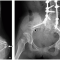
FIGURE 9.34 Tricuspid atresia. Frontal chest radiograph in cyanotic infant shows mildly decreased pulmonary vascularity, normal heart size, and concavity (arrow) in the region of main pulmonary artery.

Surgical treatment involves the establishment of pulmonary artery blood supply, which in the neonatal period is achieved by a modified BT shunt (right subclavian artery to RPA). Subsequently, during infancy after the pulmonary vascular resistance drops, the BT shunt is taken down, and a Glenn shunt/superior cavopulmonary anastomosis (SVC to RPA) is performed. Similar to HLHS, the Fontan shunt/total cavopulmonary anastomosis is performed at 18 months to 4 years of age, after which all the SVC and IVC blood returns directly to the pulmonary arteries rather than to the heart.30


Double Inlet Left Ventricle

Double inlet LV is a type of complex single ventricular connection in which there is a dominant LV, two AVVs, and a rudimentary right ventricular remnant that communicates with the LV via a small VSD (known as a bulboventricular foramen).31 Patients with this condition usually have atrial and visceral situs solitus but the ventricular looping may be anomalous. Both AVVs empty into the LV (Fig. 9.35). The great arteries are usually malposed so the left ventricular chamber gives rise to the pulmonary artery and the right ventricular chamber gives rise to the aorta, thereby placing these patients at risk of subaortic stenosis, HLHS, interrupted
aortic arch (IAA), and aortic coarctation. Pulmonary outflow tract obstruction may also occur with either concordant or discordant ventriculoarterial connections.31 Presentation is usually in the neonatal period with cyanosis and congestive heart failure.


FIGURE 9.35 Double inlet left ventricle. Axial bright blood magnetic resonance image in four-chamber plane shows both atrioventricular valves (arrows) emptying into the left ventricular chamber (LV) with atretic right ventricle (RV). The right atrium (RA) and left atrium (LA) are also connected with ostium secundum atrial septal defect (*).

Chest radiography usually demonstrates cardiomegaly, and in patients with subaortic stenosis, the pulmonary vascularity is increased. The intracardiac anatomy is usually adequately depicted with echo. If required, cardiac MRI can demonstrate both AVVs emptying into a dominant ventricular chamber with a left ventricular morphology. The rudimentary chamber may be identified as a morphologic RV by the presence of the moderator band. Cardiac MRI can also demonstrate the outflow tract relationships, the presence of outflow tract stenosis, the size of the bulboventricular foramen, and great artery anatomy. CTA may be indicated to better delineate the aortic arch anomalies in patients with aortic coarctation/interruption.

Patients with double inlet LV usually undergo a single ventricle staged surgical palliation. In patients with severe subaortic stenosis and hypoplastic aorta, a Norwood type operation is performed shortly after birth (similar to HLHS), followed by the Glenn and Fontan shunts.31


Unbalanced Atrioventricular Septal Defects

AVSDs result from failure of fusion of the endocardial cushions leading to a deficient AV septum and a common AVV rather than separate openings and attachments for the mitral and tricuspid valves.32 If this common AVV is positioned so that one ventricle receives the larger proportion of AV inflow and the other ventricle is hypoplastic, an unbalanced AVSD results. Most commonly, there is a dominant RV and hypoplastic LV with similar physiology to HLHS (Fig. 9.36).32,33 The corollary anomaly with a dominant LV and right ventricular hypoplasia results in a physiology similar to tricuspid atresia. Unbalanced AV canal defects are seen in association with Down syndrome and heterotaxy as well as isolated anomalies.32,33 Clinically affected pediatric patients usually present with cyanosis, murmur, and cardiac failure. Metabolic acidosis and peripheral ischemia may be present in the setting of HLHS-like morphology.33,34

Chest radiography demonstrates cardiomegaly, pulmonary edema, and situs anomalies in the setting of heterotaxy. Echo is used for the initial diagnosis with MRI reserved for borderline cases with less severe forms of left-sided obstruction and larger LVs that potentially could support a biventricular type cardiac repair. In these instances, cardiac MRI can accurately measure the LVEDV, screen for the presence of endocardial fibroelastosis, and assess for left ventricular outflow tract obstruction, which may help predict suitability for a biventricular repair.25 In cases of heterotaxy, CTA or MRA can accurately depict the complex systemic and pulmonary venous anomalies. Additionally, CTA or MRI can also assess the upper abdomen for concomitant splenic and GI rotation anomalies.


FIGURE 9.36 Unbalanced atrioventricular canal defect in a 2-month-old girl with heterotaxy. Bright blood magnetic resonance image in four-chamber plane shows large atrioventricular canal defect (AVC) positioned over the right ventricle (RV) (i.e., right-sided dominance) resulting in hypoplastic left ventricle (LV). Also noted is azygos continuation (AZ) of IVC. LA, left atrium; RA, right atrium.

The surgical approach depends on the morphology of the unbalanced defect. It may involve a biventricular repair in patients with milder forms and appropriate biventricular sizes, a three-stage palliation (Norwood, Glenn, Fontan) in patients with a HLHS-like morphology, or a modified BT shunt followed by Glenn and Fontan shunts in patients with a tricuspid atresia-like morphology.34


Conotruncal Anomalies

The conotruncal region of the heart comprises of the ventricular outflow tracts, the aortic and pulmonary valves, and the outlet (conal and membranous) portions of the interventricular septum. The embryologic precursors of this region are the distal bulbus cordis and truncus arteriosus. The term conotruncal anomalies encompasses TOF, pulmonary atresia with intact ventricular septum, transposition of great vessels, DORV, truncus arteriosus, and even IAA (IAA is discussed with the arch anomalies portion of this chapter). There is a high incidence of DiGeorge syndrome (chromosome 22q11.2 deletion) in patients with conotruncal anomalies.35


Tetralogy of Fallot

TOF arises from a single pathologic defect in the formation of the conus (infundibulum).36,37 In TOF, the conus is abnormally superiorly and anteriorly displaced, narrowing the RVOT, which leads to right ventricular hypertrophy. An outlet type VSD results where the conus would have formed the superior part of the VSD, and the aorta overrides the VSD (Figs. 9.37 and 9.38). The RVOT narrowing results in a spectrum of pulmonary valve pathology ranging from mild dysplasia to
pulmonary atresia. Absence of the pulmonary valve may also occur in TOF, and this entity is discussed separately later in this chapter. The variable degree of RVOT/pulmonary valve stenosis results in variations in branch pulmonary artery anatomy. In most cases, the pulmonary arteries are normal or mildly diminished in size. However, with pulmonary valve atresia, the branch pulmonary artery anatomy may be very complex. The branch pulmonary arteries may be connected to each other (confluent) and solely supplied either by the ductus arteriosus or by major arteriopulmonary collaterals (MAPCAs).38,39 Nonconfluent branch pulmonary arteries may be solely supplied by multiple MAPCAs. MAPCAs may also supply individual segments of the lung directly without going through the pulmonary arterial branches.


FIGURE 9.37 Diagram of anatomy of tetralogy of Fallot with right ventricular outflow tract (RVOT) and pulmonary valvar stenosis. The aortic valve (AoV) can be seen through the ventricular septal defect (VSD). There is RVOT stenosis in the subpulmonary valve region, hypoplastic pulmonary valve (PV), hypoplastic pulmonary arteries. A right aortic (Ao) arch is present in ˜25% of patients. LA, left atrium; LV, left ventricle; MPA, main pulmonary artery; RA, right atrium; RV, right ventricle.

Only gold members can continue reading. Log In or Register to continue

Stay updated, free articles. Join our Telegram channel

Oct 13, 2018 | Posted by in PEDIATRIC IMAGING | Comments Off on Heart

Full access? Get Clinical Tree

Get Clinical Tree app for offline access