• A chronic progressive transmural granulomatous inflammatory bowel disease • There are typically discontinuous (‘skip’) lesions with asymmetrical bowel wall involvement • It can affect any part of the GI tract – however it almost always affects the terminal ileum (in 95% of cases) • Inflammatory polyps (pseudopolyps): small, discrete round filling defects • Thickened valvulae conniventes: they can also be distorted, blunted or flattened (they are due to hyperplasia of the lymphoid tissue which causes an obstructive lymphoedema) • Fistulae formation: this can involve adjacent loops of ileum, caecum or sigmoid colon • Bowel wall sacculation: this is secondary to fibrosis within healing eccentric ulcers – NECT: the stratified appearance is due to a thickened muscularis mucosae and submucosal fatty infiltration – CECT: the stratified appearance is due to acute inflammation with submucosal oedema and enhancement of the mucosa and muscularis propria • Although any part of the GI tract can be involved, the ileum is the commonest involved site (esp. the terminal ileum and ileocaecal junction) • Discrete transverse and circumferential ulcers, mucosal fold thickening and strictures are the main radiological features • Ulcerative, hypertrophic or fibrotic forms are described: • Fleischner sign: a thickened patulous ileocaecal valve seen in conjunction with a narrowed terminal ileum • Stierlin’s sign: this is due to rapid emptying of contrast through a gaping incompetent ileocaecal valve and into a conically contracted caecum • A submucosal mesenchymal (non-epithelial) tumour appearing to arise from the muscularis propria • The commonest gastrointestinal mesenchymal tumour (< 1% of all GI tract tumours) • Location: stomach (40–70%) > small intestine (20–50%) > large intestine and rectum (5%) • 90% of tumours will express KIT (CD 117) which is a tyrosine kinase growth factor receptor • Increased prevalence of GIST with NF-1 • Unfavourable prognostic signs: tumour size >5cm • Carney’s triad: a genetic syndrome of young women
Small bowel
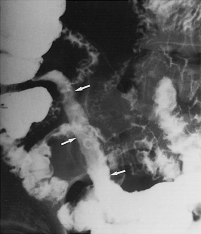
CROHN’S DISEASE (CD)
CROHN’S DISEASE
DEFINITION
RADIOLOGICAL FEATURES
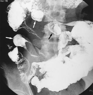
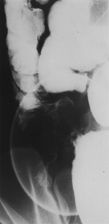
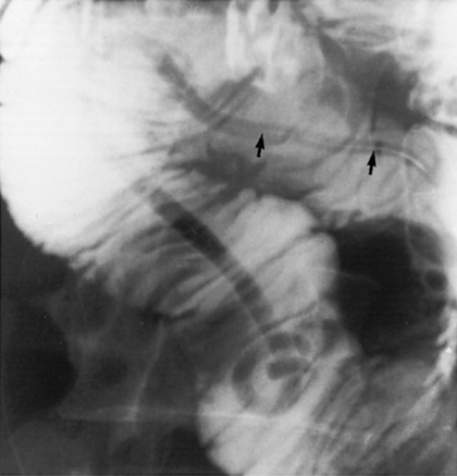
Barium studies
 Aphthoid ulcers: characteristic superficial ulcers that do not penetrate the muscularis mucosa
  they appear as small collections of barium with surrounding radiolucent oedematous margins
  en face they appear as a dense amorphous barium pool with a surrounding black halo
 Longitudinal ulcers: these run along the ileal mesenteric border
 these are not a frequent finding
 Thickened bowel wall segments will displace adjacent barium-filled loops
 ‘Skip lesions’: discontinuous involvement of the bowel wall
 These may be short, long, single or multiple (the latter is virtually diagnostic of CD)
  solitary strictures are common and may be accompanied by proximal (prestenotic) dilatation
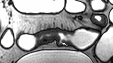
 ‘String sign’: tubular narrowing of the intestinal lumen secondary to oedema and spasm (± scarring)
 Other sites: urinary bladder
  perianal region (leading to a ‘watering can’ perineum)
  occasionally the skin and vagina
 it can also be seen with ischaemic strictures or scleroderma (with wide ‘square’-shaped diverticulae)
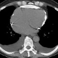
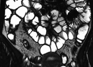
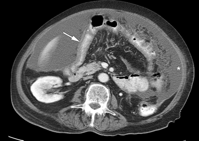
CT
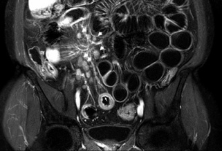
 The transmural disease leads to greater wall thickening than seen with UC
  mild reactive adenopathy (<1cm) can be present
 ‘Dirty fat’: transmural inflammation of the small bowel usually involves the adjacent mesentery
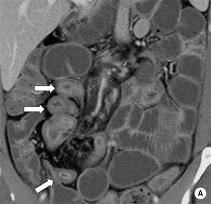
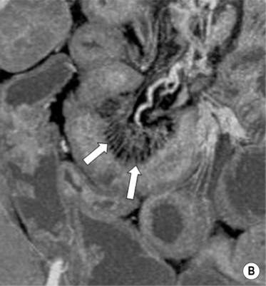
 ‘Target’ or ‘halo’ sign: a homogeneous or stratified appearance is seen on both NECT and CECT:
 ‘Creeping fat’: fat accumulates on the serosal surfaces
INFECTIONS/INFESTATIONS OF THE SMALL BOWEL
TUBERCULOSIS (TB)
Radiological features
 there are often multiple bowel lesions
Barium follow through (ileocaecal tuberculosis)
 Ulcerative: there are discrete ulcers with a ‘shaggy’ edge – these tend to be large and circumferential
GASTROINTESTINAL STROMAL TUMOURS (GISTs) AND CARCINOID TUMOURS
GASTROINTESTINAL STROMAL TUMOURS (GISTs)
Definition
 GISTs may arise from the interstitial cells of Cajal (these serve a gastric pacemaker function)
 leiomyomas and leiomyosarcomas are rare at these sites
Pearls
 this differentiates a GIST from other gastrointestinal mesenchymal tumours (e.g. leiomyoma or leiomyosarcoma)
 infiltration into adjacent organs
  metastases
  a high mitotic and proliferation index
CARCINOID TUMOURS OF THE GASTROINTESTINAL TRACT
Clinical presentation


 they may lead to abscess formation, sinuses or fistulae


 it is the most common cause of bowel loop separation in CD

 these may contain gas – this is usually due to enteric or cutaneous fistulae and sinus tracts rather than due to a gas-producing bacteria
 ulceration
  cobblestoning
  a ‘target’ appearance
  fibrofatty proliferation
  vascular engorgement
  adenopathy
  luminal narrowing and stenosis
 spondylitis
  symmetrical sacroilitis
  renal stones
  complications of steroid treatment (e.g. avascular necrosis)
  primary sclerosing cholangitis
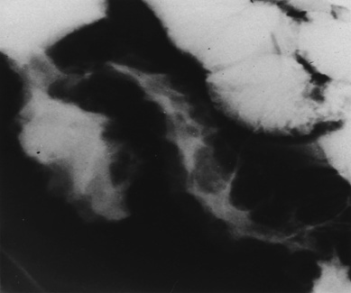

 it used to be secondary to pulmonary disease but is now more likely to be of primary bovine origin (from drinking unpasteurized milk)
 liver
liver  spleen
  lymph nodes
  peritoneum
  female genital tract
 fever
fever  weight loss
  diarrhoea
  intestinal obstruction
  bowel perforation (rare)
 it may be difficult to distinguish from lymphoma
 there may be enlarged rim enhancing low-density mesenteric nodes (due to caseous liquefaction)
 acute inflammation of the terminal ileum is often indistinguishable clinically from an acute appendicitis
 tortuous, thickened mucosal folds with small discrete nodular filling defects of lymphoid hyperplasia
  there can be mural thickening
 the appendix is the most commonly affected site
 previous surgery
  neoplasms
  diabetes
  steroids
  poor dental hygiene
 it is contracted through contaminated drinking water
 small well-defined nodular lymphoid hyperplasia in patients who also have dysgammaglobulinaemia
 thickened or absent valvulae conniventes within the duodenum and proximal jejunum
  severe cases may show a rigid ‘pipestem’ stenosis with irregular narrowing
 it may be indistinguishable from Crohn’s disease (although the mucosa remains intact)
 it is very commonly seen in the tropics
 there can be associated oesophagitis, gastritis or colitis
 thickened small bowel folds
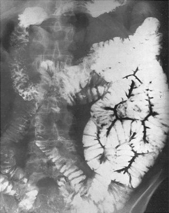


 abdominal pain
  abdominal mass
  ileus
ileus  GI bleeding
  weight loss
 the border of the smooth mucosal surface forms right or obtuse angles with the adjacent mucosa
 coarse mottled calcifications (25%)
  occasionally pedunculated (and may obstruct the pylorus or duodenum/act as a lead point for an intussusception)
  hypo- or hypervascular
 T2WI: low to intermediate SI
  T1WI + Gad: variable heterogeneous enhancement
 it is multiple in ⅓ of cases
 it can present with abdominal pain, obstruction, or an abdominal mass
  GI haemorrhage is very rare