| SKULL BASE REGION | Cerebellopontine angle/internal auditory canal |
| HISTOPATHOLOGY | N/A |
| PRIOR SURGICAL RESECTION | No |
| PERTINENT LABORATORY FINDINGS | Pretreatment audiogram: Class A hearing with bilateral sensorineural hearing loss, more pronounced in tumor ear; word recognition score at 100% in right ear and 90% in left ear |
Case description
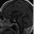
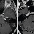
The patient presented at 71 years of age with sudden right-sided hearing loss and tinnitus. Magnetic resonance imaging (MRI) with gadolinium contrast was obtained, which revealed a small enhancing intracanalicular mass consistent with vestibular schwannoma (VS). The tumor, retrospectively, had been present on another MRI obtained the previous year as part of a research study ( Figure 8.40.1 ). An audiogram revealed a drop-off in right-sided hearing above 1000 Hz, with maintained word recognition at 100% ( Figure 8.40.2 ). Due to the small size of the tumor and good hearing status, he underwent stereotactic radiosurgery (SRS) for the tumor ( Figure 8.40.3 ). Serial imaging following initial SRS revealed interval growth of the tumor at 5 years ( Figure 8.40.4 ). Hearing decreased ( Figure 8.40.5 ) but was improved with hearing aids, and he developed imbalance. He underwent repeat Gamma Knife radiosurgery (GKRS) ( Figure 8.40.6 ).
| Radiosurgery Machine | Gamma Knife – Perfexion |
| Radiosurgery Dose (Gy) |
|
| Number of Fractions |
|


Related posts:
 Esthesioneuroblastoma – delayed postoperative radiosurgery for recurrence at long-term
 Null cell – delayed postoperative radiosurgery for growing perioptic residual
 Suprasellar non-small cell lung carcinoma metastasis – upfront radiosurgery
 Chondrosarcoma – definitive radiosurgery after subtotal resections
 Large vestibular schwannoma – delayed postoperative radiosurgery for growing residual
 Capillary hemangioma – postoperative radiosurgery for residual tumor
Stay updated, free articles. Join our Telegram channel

Full access? Get Clinical Tree


