Abstract
Smith-Lemli-Opitz syndrome (SLOS) is an autosomal recessive disorder caused by mutations in the gene encoding the 7-dehydrocholesterol reductase (7-DHC) resulting in decreased or absent function of this enzyme responsible for catalyzing the final step in cholesterol synthesis. Increased levels of 7-DHC and typically a reduction in cholesterol synthesis are seen in affected individuals. Diagnosis is made based on elevated 7-DHC. The estimated incidence of SLOS is 1 : 39,000 conceptions, with a carrier rate of approximately 1%. Higher carrier prevalence is reported in individuals of European or North American heritage. SLOS is characterized by growth failure, cognitive delay, behavioral disturbances, dysmorphic faces, and congenital defects. There is marked variability in the presentation of this condition ranging from mild effects that an individual remains undiagnosed, to fetal or neonatal death. The following findings on ultrasound (syndactyly/polydactyly, growth restriction, microcephaly, micrognathia, cleft palate, cardiac defects, and ambiguous male genitalia) in association with low maternal serum estriol and normal karyotype are highly suggestive of SLOS. Prenatal testing is available in at-risk pregnancies. Common postnatal therapies for SLOS include dietary cholesterol supplementation and surgical intervention, for the given defect or defects as appropriate.
Keywords
SLOS, Smith-Lemli-Opitz syndrome, SLO syndrome, RSH syndrome
Introduction
Smith-Lemli-Opitz syndrome (SLOS) is an autosomal recessive disorder caused by abnormal cholesterol synthesis. SLOS was originally named RSH syndrome, derived from the surnames of the first three families identified with this disorder. SLOS is characterized by growth failure, cognitive delay, behavioral disturbances, dysmorphic faces, and congenital malformations. There is marked variability in presentation of this condition among affected individuals.
Although SLOS was historically divided into two subtypes, with type I being milder and type II being more severe, there is now thought to be a clinical and biochemical continuum. SLOS is caused by mutations in the gene encoding the 7-dehydrocholesterol reductase (DHCR7 gene). These mutations results in elevation of the cholesterol precursor 7-dehydrocholesterol (7-DHC) and typically decreased cholesterol synthesis. Genotype-phenotype correlations have been noted, wherein mutations that lead to lower or absent gene function are found in the most severely affected individuals and mutations that lead to more residual function are found in less severely affected individuals.
Disorder
Definition
SLOS is an autosomal recessive syndrome that is biochemically defined. Although there are abnormalities commonly associated with SLOS, diagnosis is made based on elevated 7-DHC because of the variable presentation in affected individuals. Decreased serum cholesterol is also noted in most affected individuals; however, 10% have normal serum cholesterol. Therefore the cholesterol level alone is inadequate to confirm the diagnosis.
Prevalence and Epidemiology
The estimated incidence of SLOS is 1 : 39,000 conceptions, with a carrier rate of approximately 1%. Although SLOS is panethnic, higher carrier prevalence is reported in individuals of European or North American heritage (1%–2%). Older studies identified a greater than 2 : 1 ratio for males to females affected with SLOS; however, this is likely caused by ascertainment bias. Males are often identified owing to abnormal external genitalia, a finding lacking in affected females. Recent studies demonstrate an equivalent male-to-female ratio.
Etiology and Pathophysiology
SLOS is an inborn error of metabolism syndrome caused by homozygous mutations in the DHCR7 gene, which codes for the enzyme 7-DHC reductase. These mutations lead to decreased or absent function of this enzyme. Seven-DHC reductase is responsible for catalyzing the final step in cholesterol synthesis, wherein 7-DHC is converted to cholesterol. Increased levels of 7-DHC are seen in affected individuals. The clinical sequelae of SLOS may be secondary to one or all of these factors: low levels of cholesterol, deficient total sterols, and toxic effects of markedly elevated concentrations of 7-DHC.
The severity of the condition is likely related to the percent of remaining 7-DHC activity in the individual. Cholesterol has many functions in the body and is essential for fetal development. It is a required precursor to many important molecules, such as androgens. Low or absent cholesterol explains the incidence of abnormal or feminized external genitalia in XY individuals with SLOS. The other abnormalities noted in SLOS are likely caused by the fact that cholesterol interacts with “hedgehog” proteins, which encode inductive signals during embryogenesis. If an individual has deficient cholesterol levels, it may be caused by downregulation of hedgehog proteins. The spectrum of anomalies noted in SLOS is consistent with anomalies in individuals with mutations in these hedgehog proteins.
In contrast to many other metabolites, minimal cholesterol is supplied to the developing fetus by the mother. Signs of SLOS are often noted prenatally because cholesterol is an essential building block during fetal development. For this reason, SLOS may be suspected if maternal estriol levels are low during second-trimester maternal serum screening, particularly when characteristic congenital anomalies are also noted on ultrasound (US). Data suggest that fetuses with SLOS are likely to have low maternal serum estriol levels, low beta-human chorionic gonadotropin levels, and low alpha-fetoprotein levels. The detection rate for fetuses affected with SLOS by this screening method has not been established. Nonetheless, evaluation for SLOS is commonly discussed if the maternal serum estriol level is lower than 0.3 multiples of the median adjusted for gestational age.
Manifestations of Disease
Clinical Presentation
SLOS presentation can range from such mild effects that an individual remains undiagnosed to serious congenital anomalies, cognitive delay, and fetal or neonatal death. Findings commonly associated with SLOS are listed subsequently :
- •
general
- •
cognitive delays
- •
developmental delays
- •
prenatal and postnatal growth restriction
- •
- •
behavioral disturbances
- •
autism spectrum disorder
- •
self-injury
- •
- •
central nervous system malformations
- •
holoprosencephaly
- •
agenesis or partial agenesis of corpus callosum
- •
- •
craniofacial
- •
microcephaly
- •
cleft palate/arched palate/bifid uvula
- •
dysmorphic facial features (anteverted nares, micrognathia, blepharoptosis)
- •
cataracts
- •
- •
extremities
- •
syndactyly of toes two and three
- •
polydactyly
- •
- •
cardiac defects (septal defects)
- •
renal hypoplasia
- •
hypoplastic or unilobar lungs
- •
visceral anomalies
- •
abnormal external genitalia or cryptorchidism in XY individuals
- •
photosensitivity
Imaging Technique and Findings
Ultrasound.
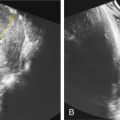
The following findings on US in association with low maternal serum estriol are highly suggestive of SLOS ( Figs. 145.1–145.4 ) :
- •
syndactyly/polydactyly
- •
growth restriction
- •
microcephaly
- •
micrognathia
- •
cleft palate
- •
cardiac defects


