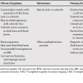
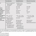
104 Because the differential diagnosis of solid peritoneal masses is very broad, it is useful to divide the differential in masses that diffusely involve the peritoneal space and those that are confined to the mesentery. The most likely etiology is peritoneal carcinomatosis, most commonly from an ovarian, colon, or stomach primary site. This is often seen in conjunction with ascites. There are many other possible etiologies, which are rare and overlap in appearance. Peritoneal tuberculosis can resemble peritoneal carcinomatosis. Certain imaging finding are helpful in differentiating tuberculous peritonitis from peritoneal carcinomatosis (Table 104.1). Peritoneal lymphomatosis is rare and usually cannot be distinguished from peritoneal carcinomatosis. Ascites with loculation or septation and a diffuse distribution of enlarged lymph nodes may suggest the diagnosis.1 The gastrointestinal tract is the most common primary site in peritoneal dissemination of leiomyosarcomas, but other primary sites such as the mesentery, retroperitoneum, genitourinary tract and soft tissues are also possible. Features suggesting peritoneal leiomyosarcomatosis are necrosis, liver metastases, and minimal or absent ascites.2
Solid Peritoneal Masses
Diffuse Peritoneal Masses
Peritoneal Tuberculosis
Peritoneal Lymphomatosis
Peritoneal Leiomyosarcomatosis
| Carcinomatosis | Tuberculosis | |
|---|---|---|
| Irregular peritoneal infiltration | More common | Less common |
| Mesenteric Changes | Less common | More common |
| Macronodules >5 mm in diameter | Less common | More common |
| Low density center in masses* | Less common | More common |
| Calcification† | Less common | More common |
| Thin omental line covering infiltrated omentum | Less common | More common |
* Can be seen in treated lymphoma, Burkitt’s lymphoma, metastases, abscess, and Whipple’s disease
† Can be seen in implants from ovarian serous adenocarcinoma
Peritoneal Amyloidosis
Peritoneal amyloidosis is a rare manifestation of systemic amyloidosis. Calcification and poor or absent enhancement of the amyloid deposits may be helpful in suggesting the diagnosis.3
Peritoneal Sarcoidosis
Peritoneal sarcoidosis is rare and usually presents with ascites.4 However, ascites in the setting of sarcoidosis is more likely related to cardiac or hepatic disease rather than peritoneal involvement.5
Desmoplastic Small Round Cell Tumor
Stay updated, free articles. Join our Telegram channel

Full access? Get Clinical Tree


