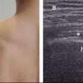
Figure 10-2. Pectoralis major tendon. [A] Positioning of the probe. [B] Corresponding 12-5 MHz US image depicts the typical fibrillar appearance of the tendon (arrowheads). Gt= greater tuberosity. Lt= lesser tuberosity. Delt= deltoid.
Related posts:

Stay updated, free articles. Join our Telegram channel

Full access? Get Clinical Tree


