SPECT-CT Imaging of Sentinel Lymph Nodes in Breast Cancer
Daniela B. Husarik
Hans C. Steinert
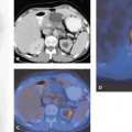
One of the promising clinical uses of integrated single-photon emission computed tomography (SPECT) and computed tomography (CT) in breast cancer is in the detection of the sentinel lymph node(s) (SLN). Tomography rather than planar imaging is better able to distinguish a SLN close to the injection site, and integrated imaging defines the anatomical location of the SLN to better advantage for the surgeon. Although the literature on this technique is still sparse, we at our institution exclusively use SPECT-CT for SLN detection, since this technique has become available and early results indicate that the procedure is superior to planar and SPECT-only imaging (see Fig. 46.1).
Rationale
The most common site of regional metastasis of breast cancer is the axillary lymph nodes. Additional drainage sites are supraclavicular, internal mammary, intramammary, and interpectoral nodes. The status of lymph nodes is the most important prognostic variable in the staging of breast cancer. Patients with histologically negative axillary lymph nodes have significantly better survival rates than patients with positive nodes. Clinical evaluation for predicting nodal disease is inadequate.
Lymphatic mapping of the sentinel lymph node (SLN) with radiolabeled colloid is a promising technique to investigate the drainage nodal basins of breast cancer as well as other cancers. The lymphatic mapping is followed by SLN biopsy for the histologic examination and detection of microscopic metastases. In many cases, particularly in the United States, SLN localization and sampling have eliminated many full axillary lymph node dissections. In breast cancer, SLN biopsy has the great advantage of being a minimally invasive procedure. Therefore, the risk of lymphedema, which often occurs after axillary lymph node dissection, is minimized. With the SLN information, full axillary lymph node dissections can be restricted only to patients who show microscopic lymph node involvement.
To facilitate a minimally invasive biopsy, an accurate localization of SLN is necessary. For localization of SLN, the current classification system of the American Joint Committee (AJCC) is used. The lymph nodes are divided into low axillary (level I); mid-axillary (level II); high axillary, apical (level III); supraclavicular; and internal mammary nodal stations.
Until now, planar images have been used for SLN imaging. However, limitations of planar images for the detection of SLN in breast cancer are well known. SLN close to the injection site can be overlooked and may result in false-negative cases. The exact anatomical localization of the SLN is difficult using planar as well as single-photon emission computed tomography (SPECT) images. These weak points of planar imaging can be overcome by integrated
SPECT and computed tomography (CT) scanning, combining the benefits of SPECT and the anatomical information of CT.
SPECT and computed tomography (CT) scanning, combining the benefits of SPECT and the anatomical information of CT.
Stay updated, free articles. Join our Telegram channel

Full access? Get Clinical Tree


