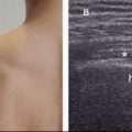
Figure 8-2. Thickening of the proximal coracohumeral ligament. [A] Positioning of the probe. [B] Corresponding 12-5 MHz US image depicts hypoechoic thickening of the coracohumeral ligament (arrowheads). This finding is consistent with the clinical diagnosis of adhesive capsulitis. [C] Comparative contralateral image shows normal ligament in asymptomatic shoulder (arrowheads). Cp= coracoid process.

Figure 8-3. Thickening of the distal coracohumeral ligament at the rotator interval. [A] Positioning of the probe. [B] Corresponding 12-5 MHz US image depicts hypoechoic thickening of the coracohumeral ligament (arrowheads). [C] Comparative contralateral image demonstrates the normal coracohumeral ligament in asymptomatic shoulder. T= long head of the biceps brachii tendon. Hum= humerus.

Figure 8-4. Hypoechoic material surrounding the long head of the biceps brachii tendon at the rotator interval. [A] Positioning of the probe. [B] Corresponding 12-5 MHz US image depicts thickened and hypoechoic material (asterisks) surrounding the long head of the biceps brachii tendon (t). This finding is not specific for adhesive capsulitis and may also be identified in pulley lesion caused by degenerative changes, acute trauma, repetitive microtrauma, or injury associated with a rotator cuff tear. The rotator interval contains the coracohumeral and superior glenohumeral ligaments, which are both encased in a capsulosynovial membrane and subject to the same pathological processes as the remainder of the glenohumeral joint. Hum= humerus.

Figure 8-5. Increased vascularity around the long head of the biceps brachii tendon at the rotator interval. [A] Positioning of the probe. [B] Corresponding 12-5 MHz US image depicts increased vascularity at the rotator interval during freezing stage. T= long head of the biceps brachii tendon. Hum= humerus.
Stay updated, free articles. Join our Telegram channel

Full access? Get Clinical Tree


