Sphenopetroclival meningioma – delayed postoperative radiosurgery for growing residual
SKULL BASE REGION
Sphenopetroclival
HISTOPATHOLOGY
Meningioma, transitional
PRIOR SURGICAL RESECTION
Yes
PERTINENT LABORATORY FINDINGS
N/A
Case description
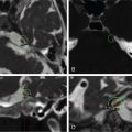
The patient is a 46-year-old female who presented to another hospital with progressive headache, visual loss, dysphagia, dizziness, and gait disturbances. Brain magnetic resonance imaging (MRI) showed a massive petroclival meningioma with obstructive hydrocephalus ( Figure 7.30.1 ), necessitating placement of a ventriculoperitoneal shunt. Symptoms persisted, and she was referred to our institution. She underwent resection via a retrosigmoid approach, leaving a tumor remnant in Meckel’s cave and the cavernous sinus ( Figure 7.30.2 ). Postoperative course was uneventful, and patient recovered her full function after a short period of rehabilitation. Follow-up MRI at 1 year revealed a slight increase in size of the tumor remnant ( Figure 7.30.3 ), and stereotactic radiosurgery (SRS) was recommended to stop tumor growth ( Figure 7.30.4 ).
Radiosurgery Machine
CyberKnife
Radiosurgery Dose (Gy)
25, at the 78% isodose line
Number of Fractions
5
Figure 7.30.1.
Preoperative postcontrast T1-weighted image showing a large sphenopetroclival mass.
Figure 7.30.2.
Postoperative postcontrast T1-weighted images showing a tumor remnant in Meckel’s cave and the left cavernous sinus.
Figure 7.30.3.
Follow-up images at 1 year after resection showing increase in size of the tumor remnant.
Figure 7.30.4.
Imaging of the treatment plan.
Critical Structure
Dose Tolerance
Chiasm
25/27 Gy, 4/5 fractions
Optic nerve
25/27 Gy, 4/5 fractions
Oculomotor nerves (III, IV, VI)
25/27 Gy, 4/5 fractions
Trigeminal nerve
N/A
Only gold members can continue reading. Log In or Register to continue
Apr 6, 2024 | Posted by drzezo in GENERAL RADIOLOGY | Comments Off on Sphenopetroclival meningioma – delayed postoperative radiosurgery for growing residual


 Esthesioneuroblastoma – delayed postoperative radiosurgery for recurrence at long-term
 Null cell – delayed postoperative radiosurgery for growing perioptic residual
 Chondrosarcoma – definitive radiosurgery after subtotal resections
 Trigeminal neuralgia due to microvascular conflict – upfront radiosurgery
 Capillary hemangioma – postoperative radiosurgery for residual tumor
 Superior sagittal sinus meningioma – delayed postoperative, multisession radiosurgery for growing residual


