Clinical Presentation
The patient is a 70-year-old woman with a long history of progressive neck pain and bilateral shoulder pain, right more than left, and right arm numbness. She has a mild gait disturbance requiring use of a cane. She underwent right rotator cuff surgery 7 months prior, with no improvement in symptoms. In fact, the neck and right shoulder pain has progressively worsened since the surgery. She has moderate right deltoid and biceps weakness on physical examination.
Imaging Presentation
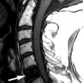
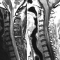
Magnetic resonance (MR) imaging reveals an ovoid lesion in the posterior portion of the spinal cord at the C4-5 level associated with mild cord enlargement ( Fig. 67-1 ) . The lesion is T2 hyperintense centrally and surrounded by a well-defined region of T2 hypointensity ( Figs. 67-1 and 67-2 ) . The lesion is slightly hyperintense at its periphery on T1-weighted images ( Fig. 67-3 ) and displays subtle enhancement after administration of IV paramagnetic contrast agent ( Fig. 67-4 ) . The imaging appearance is classic for a spinal cord cavernous angioma (cavernoma).


Discussion
Cavernous malformations are also called cavernous malformations, cavernous hemangiomas , cavernomas, “cryptic” malformations, or angiographically occult vascular malformations . Cavernous angiomas are uncommon spinal cord lesions, comprising 5% of intramedullary cord lesions. These lesions commonly occur in the brain and much less commonly in the spinal cord. Half of the cases arise in the cervical cord and the other half in the thoracic cord ( Figs. 67-5 to 67-7 ) , or in the conus medullaris. They are more common in females and usually manifest in young to middle-aged adults. Cavernous angiomas can be isolated lesions or can be one of multiple lesions occurring in the brain and spinal cord. Up to 40% of patients with spinal cord cavernous angiomas have at least one brain cavernous angioma. Multiple brain and cord cavernous angiomas may occur sporadically or can be associated with a familial syndrome that is autosomal dominant with variable penetrance in a minority of cases. In familial cases, spinal cord cavernomas may coexist with cavernomas in the brain, retina, and skin as well as cavernous hemangiomas in the vertebral bodies. Cavernous angiomas manifesting in the pediatric patient are more likely to manifest with acute symptoms, more likely to hemorrhage, and more likely to be familial. Up to 50% of pediatric patients with spinal cord cavernomas have associated hemorrhage. Rarely, spinal cord angiomas can occur in patients with Klippel-Trénaunay-Weber syndrome.