A. PET/CT scan
B. Percutaneous biopsy
C. Antifungal treatment
D. No further workup or treatment
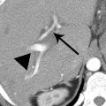
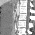
2 In patients 40 years or older, what is the most common etiology of the splenic finding on the following CT image?

A. Sickle cell disease
B. Embolic disease
C. Pancreatitis
D. Splenic torsion
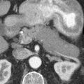
3 The following CT image is from a patient with a history of an orthotopic liver transplant. What is the most common organism that causes these splenic and hepatic findings?

A. Candida species
B. Mycobacterium avium complex
C. Bartonella henselae
D. Pneumocystis jiroveci
4a A 38-year-old woman presents for evaluation of diffuse abdominal pain, bloating, and anorexia. The patient has a history of surgery after a fall as a child. Two images from a CT scan are shown. Which is the most appropriate test for confirmation of the suspected diagnosis?

A. MRI without and with contrast
B. PET/CT
C. Sulfur colloid scan
D. Hepatobiliary iminodiacetic acid (HIDA) scan
4b A Tc-99m sulfur colloid scan was performed. Based on the following image from the scan, what is the diagnosis?

A. Peritoneal carcinomatosis
B. Splenosis
C. Tuberculosis
D. Lymphoma
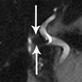
5 A 38-year-old man presenting with abdominal pain underwent an MRI to further evaluate a finding in the left upper quadrant seen on ultrasound. What is the most likely diagnosis?

Top row: T2W and T1W. Bottom row: arterial and venous phase FS T1W+gad.
A. Pancreatic adenocarcinoma
B. Splenule
C. Peritoneal carcinomatosis
D. Aneurysm
6 A 66-year-old woman presents with a palpable neck mass but is otherwise asymptomatic. A CT scan was performed. What is the most likely explanation for the appearance of the spleen?

A. Gaucher disease
B. Normal early enhancement pattern
C. Lymphoma
D. Blunt trauma
7 A 47-year-old man with alcoholic pancreatitis and acute intoxication fell against a table. At the time of transfer from outside hospital, hematocrit was 29%, systolic blood pressure was 116 mm Hg, and heart rate was 91 bpm. The patient was managed conservatively with supportive care. Twenty-four hours later, the hematocrit level was 25%, systolic blood pressure was 82 mm Hg, and heart rate was 130 bpm. A CT scan was performed as shown below. What is the most appropriate next step in management of the splenic findings?

A. Selective transcatheter arterial embolization
B. Percutaneous drainage
C. Surgery
D. Continuation of supportive care without intervention
8 A 59-year-old man with a history of melanoma presents for routine surveillance imaging. A CT was performed with image shown below. What is the most likely diagnosis of the finding in the spleen?

A. Littoral cell angiomas
B. Lymphangiomas
C. Pyogenic abscesses
D. Metastases
9 The findings in the spleen on the following MR images on a 1.5-T magnet are most commonly associated with which disease process?

T1W in-phase, T1W out-of-phase, and T2W.
A. Myelofibrosis
B. Lymphoma
C. Portal hypertension
D. Hydatid disease
10 A 55-year-old man with known head and neck cancer presents for staging evaluation. An abdominal MRI examination was performed. What is the most likely diagnosis of the splenic finding?

Top row: axial T1W and fat-saturated T2W. Bottom row: arterial phase and delayed phase.
A. Lymphangioma
B. Metastasis
C. Hamartoma
D. Angiosarcoma
11 Patients with this appearance of the spleen as demonstrated on the following CT image are at risk for:

A. Obstructive uropathy
B. Wandering spleen
C. Splenic rupture
D. Pancreatic necrosis
12 An axial CT image of a patient with splenomegaly is shown. Which of the following is the most effective technique for reducing the artifact seen across the lower half of the image?

A. Using sequential (axial) scanning technique rather than helical scanning technique
B. Using thinner acquisition section width
C. Raising the patient’s arms
D. Asking the patient to suspend respiration
13 A patient with limb hemihypertrophy and port wine stains underwent an abdominal CT scan shown below. Which of the following syndromes does this patient have?

A. Klippel-Trenaunay syndrome
B. Von Hippel-Lindau syndrome
C. Beckwith-Wiedemann syndrome
D. Neurofibromatosis type I

A. Situs solitus
B. Situs inversus
C. Asplenia
D. Polysplenia
15 Match the splenic imaging findings in patients 1 to 3 with the most likely diagnosis A to C. Each option may be used only once.
A. Sickle cell anemia
B. Posttraumatic sequela
C. Old healed granulomatous infection
Patient 1. A 74-year-old woman with CT and ultrasound images.

Patient 2. A 26-year-old man.


Patient 3. A 52-year-old woman.
16 A 43-year-old man with no significant past medical history presents with upper abdominal pain. The patient was afebrile with a normal white blood cell count. An image from a contrast-enhanced CT scan and coronal image from a PET/CT scan are shown. Which diagnosis is most consistent with the imaging findings and clinical scenario?

A. Angiosarcoma
B. Leukemia
C. Multiple lymphangiomas
D. Multiple hemangiomas
ANSWERS AND EXPLANATIONS
1 Answer D.This lesion has imaging characteristics most consistent with a splenic hemangioma. There is a rim of enhancement in the arterial phase with centripetal fill-in in the venous phase, and enhancement remains greater than surrounding splenic tissue. This was an isolated incidental finding unrelated to the symptoms. In this setting, no further workup is required.
Overall, most splenic lesions are benign. Splenic hemangioma is the most common benign primary neoplasm of the spleen. Suspicion for malignancy and active infection is low in this setting, so PET/CT, percutaneous biopsy, and antifungal treatment are not indicated. Patients with fungal microabscesses are immunocompromised and have multiple subcentimeter lesions. Microabscesses are usually identified as hypodense lesions relative to spleen on CT, although some can show early ring or central enhancement better appreciated on MRI.
Splenic hemangiomas show a wider spectrum of appearances than hepatic hemangiomas do. Most splenic hemangiomas do not show the peripheral nodular discontinuous enhancement classically seen with hepatic cavernous hemangiomas. Lesions range from solid to cystic with variable internal complexity and enhancement. However, hemangiomas tend to be echogenic on ultrasound and on T2W MRI sequences, tend to be brighter than other splenic lesions.
References: Luna A, Ribes R, Caro P, et al. MRI of focal splenic lesions without and with dynamic gadolinium enhancement. AJR Am J Roentgenol 2006;186(6):1533–1547.
Vos PM, Barnard SA, Cooperberg PL. Chapter 105: Benign and malignant lesions of the spleen. In: Gore RM, Levine MS (eds). Textbook of gastrointestinal radiology, 4th ed. Philadelphia, PA: Elsevier/Saunders, 2015:1923–1964.
2 Answer B.Contrast-enhanced CT shows a geographic wedge-shaped area of hypoenhancement of the spleen extending from the hilum with the base toward the capsule. This finding is classic for a splenic infarct. Splenic infarcts may have an atypical round or mottled appearance or involve the entire spleen. A “rim” sign may be seen, with a thin line representing sparing of the capsule due to separate vascular supply from capsular arteries. The margins of an infarct usually become better defined over time and evolve into a residual scar with capsular retraction.
The most common cause in patients 40 years or older is embolic disease such as atrial fibrillation and endocarditis. The most common cause in patients younger than 40 years is a hematologic disorder such as sickle cell disease and leukemia. Other causes of infarcts include thrombosis or aneurysm of the splenic vessels, splenomegaly, blunt trauma, and collagen vascular disease. Splenic torsion is an uncommon phenomenon seen in patients with wandering spleen due to a long vascular pedicle and underdeveloped ligamentous attachments.
References: Rabushka LS, Kawashima A, Fishman EK. Imaging of the spleen: CT with supplemental MR examination. Radiographics 1994;14(2):307–332.
Vos PM, Barnard SA, Cooperberg PL. Chapter 105: Benign and malignant lesions of the spleen. In: Gore RM, Levine MS (eds). Textbook of gastrointestinal radiology, 4th ed. Philadelphia, PA: Elsevier/Saunders, 2015:1923–1964.
3 Answer A.This immunocompromised patient has a disseminated opportunistic fungal infection with multiple tiny hypodensities representing microabscesses in the spleen and liver. Microabscesses are usually fungal in etiology, most commonly Candida, Aspergillus, and Cryptococcus species. Microabscesses may be too small to visualize on any imaging modality, although MRI may be more sensitive than CT. On MRI, the lesions are T2 hyperintense and can show ring or central enhancement.
Bacterial abscesses that occur in the spleen tend to be larger and are most often caused by septic emboli. Clinical history is important for suggesting the diagnosis of infection since other benign and malignant processes may present with multiple small hypoattenuating lesions in the spleen, including hemangiomas, lymphangiomas, sarcoid, lymphoma, and metastases.
All of the other answer choices are infectious causes of multiple splenic hypodensities but less common. Mycobacterium avium complex (a group of atypical mycobacteria) and Pneumocystis jiroveci (a fungus) affect immunocompromised patients. Bartonella henselae is the bacterium responsible for cat-scratch disease. Patients with cat-scratch disease are usually young, and hepatosplenic involvement is uncommon. In immunocompromised patients, especially those with AIDS, Bartonella species can cause bacillary angiomatosis, a condition with multiple vascular peliosis-type lesions in the liver as well as the spleen. Treatment of these various disseminated infections is a drug regimen targeting the underlying organism.
References: Karlo CA, Stolzmann P, Do RK, et al. Computed tomography of the spleen: how to interpret the hypodense lesion. Insights Imaging 2013;4(1):65–76.
Vos PM, Barnard SA, Cooperberg PL. Chapter 105: Benign and malignant lesions of the spleen. In: Gore RM, Levine MS (eds). Textbook of gastrointestinal radiology, 4th ed. Philadelphia, PA: Elsevier/Saunders, 2015:1923–1964.
Related posts:
Stay updated, free articles. Join our Telegram channel

Full access? Get Clinical Tree


