Clinical Presentation
The patient is a 73-year-old woman with a chief complaint of low back pain. The patient states that the pain began 3 years ago with some back spasms that started while she was sitting down. There was no significant trauma at that time. The frequency of her pain progressed and for the past year she feels it has been present daily. The pain is of a deep, aching quality, severe at times, in the midline in the lower back. It is relieved by heating pads and a mild oral narcotic and made worse with attempts at dressing and when she wears low-heeled shoes. The patient is worse when sitting and somewhat better walking and standing. There is also relief when she leans over the cart at the grocery store. The pain is not intensified by coughing, sneezing, or bearing down. The patient also complains of pain in the lower extremities, slightly greater on the left side. She describes the pain as being at the lateral aspect of the thigh and the lateral aspect of the leg. The character of her leg pain is burning and sharp. There is no bowel or bladder dysfunction.
Imaging Presentation
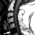
Sagittal and axial T1-weighted images demonstrate anterior subluxation of L4 relative to L5 with severe spinal stenosis secondary to the pseudo broad-based disc bulging from the anterolisthesis and facet degenerative changes. There is approximately 25% uncovering of the L4-5 disc compatible with a grade 1 spondylolisthesis ( Fig. 73-1 ) .

Discussion
Degenerative spondylolisthesis is defined as the slipping of one vertebral body with respect to an adjacent vertebral body with an intact neural arch. Generally, the direction of slip is defined as the more cephalad vertebral body in relation to the adjacent, inferior vertebral body. Anterolisthesis is defined as anterior slipping of the cephalad vertebral body in relation to the inferior one. Retrolisthesis is posterior slipping of the cephalad vertebral body in relation to the inferior one. One may also refer to lateral listhesis when there is slippage of a vertebral body left or right compared with the inferior vertebral body, which is readily seen with coronal reformatted images.
The various types of spondylolisthesis are divided into six subgroups: degenerative, isthmic, congenital/dysplastic, traumatic, pathologic, and iatrogenic. The isthmic lytic lesion of the pars interarticularis is the most common cause of spondylolisthesis and is called spondylolysis . The pars defect is what differentiates degenerative spondylolisthesis from isthmic spondylolisthesis/spondylolysis. See Chapter 69 for further discussion of spondylolysis.
Spondylolisthesis is a condition unique to humans. It is thought that spondylolisthesis developed in response to man’s ability to maintain an erect posture and the development of the lumbar lordosis. The lumbar lordosis is unique to human beings.
Degenerative spondylolisthesis usually affects people over 50 years of age. An autopsy study demonstrated the incidence of degenerative spondylolisthesis to be 4.1%, with the most affected level being L4-5, which differs from spondylolysis in which the L5-S1 level is most affected from an L5 pars defect. Most studies demonstrate a female predominance of degenerative spondylolisthesis, which has been postulated to be caused by the increased ligamentous laxity in women compared with that in men. Other potential risk factors include pregnancy, African-American ethnicity, sagittally oriented facet joints, and hyperlordosis. Degenerative spondylolisthesis can also occur in the cervical spine and has been reported in the thoracic spine as well.
Degenerative slippage occurs because of disc degenerative changes leading to facet arthritis, laxity of the paravertebral ligaments, and ineffective paravertebral muscular stabilization. The typical signs and symptoms of degenerative spondylolisthesis are often related to the degenerated and subluxed facet joints, the instability of the facet joint capsule from the ligamentous laxity, and spinal stenosis and/or neural foraminal stenosis from the slippage. Therefore, the patients often present with back/neck pain and/or radicular pain. Given that L4-5 is the most affected level, low back pain along with pain in the L5 distribution (pain in the hips with radiation down the posterolateral legs) is commonly found. The pain may be unilateral or bilateral, and the patients will often describe pain that shifts from one extremity to the other. Another common pain presentation is that of neurogenic claudication from the spinal stenosis. Spinal stenosis symptoms are present in 42% to 82% of patients who seek help because of their spondylolisthesis. Other complaints include cold feet, altered gait, and drop episodes (where the patient unexpectedly falls while walking). Like any of the conditions that cause stenosis, pain relief is often achieved by the patient with spinal flexion. This is thought to be due to an increase in the anteroposterior dimension of the spinal canal that occurs during flexion.
The natural history of degenerative spondylolisthesis has a favorable prognosis. In a 10-year follow-up study, 34% of patients who presented with degenerative spondylolisthesis had further slipping at the abnormal segment. However, 76% of patients that had degenerative spondylolisthesis without neurologic deficit remained without neurologic deficit. Eighty-three percent of the patients who had a neurologic deficit at the presentation of their degenerative spondylolisthesis and refused surgery had progression of their deficit. These patients had a poor prognosis. Overall, only 10% to 15% of patients with degenerative spondylolisthesis eventually require surgery.
Imaging Features
Spondylolisthesis can be readily visualized with plain radiographs. The lateral radiograph is most important and demonstrates anterior or posterior displacement of one vertebral body in comparison to an adjacent vertebral body ( Fig. 73-2 ) . Oblique radiographs can often identify an intact par interarticularis (the portion of bone that connects the superior and inferior articular processes of a vertebral segment) ( Fig. 73-3 ) . Lateral flexion and extension radiographs are important to evaluate for any dynamic motion of the abnormal segment and to evaluate for reduction of the spondylolisthesis. With degenerative spondylolisthesis, there may be narrowing of the disc space, hypertrophic facet degeneration, osteophytes, vacuum disc phenomenon, and/or endplate sclerosis. Computed tomography (CT) using a bone window or bone algorithm is more sensitive in evaluating the cause of the slip, in evaluating the degenerative changes, and to exclude a pars interarticularis defect ( Fig. 73-4 ) . With soft tissue algorithm CT, one is better able to evaluate the effect of the slip in relation to spinal canal narrowing, neural foraminal impingement, and lateral recess stenosis ( Fig. 73-5 ) . Magnetic resonance imaging (MRI) has largely replaced CT in the evaluation of neurologic symptoms related to the spine. MRI is more sensitive than CT in evaluating the spinal cord and cauda equina, but is less sensitive in the evaluation of the osteolytic defect of spondylolysis. In larger patients, MRI may be able to assess central canal and foraminal stenosis better than CT due to limitations of CT in large patients, particularly in the lower lumbar spine ( Figs. 73-1 and 73-6 ) .

Related posts:


Stay updated, free articles. Join our Telegram channel

Full access? Get Clinical Tree


