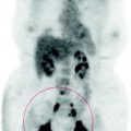
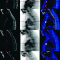
Fig. 47.1
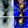
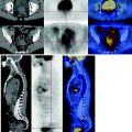
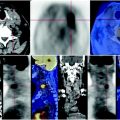
CT shows that the structural alteration is of benign type
No areas of abnormal FDG deposition in other parts of the body examined.
47.4 Conclusions
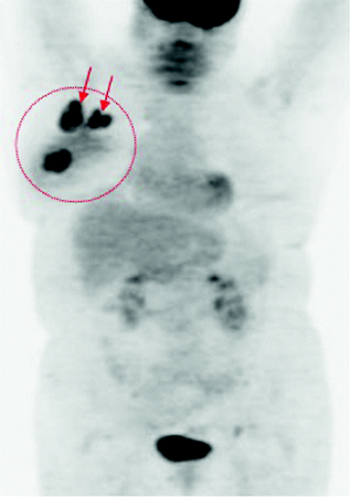
The PET scan shows a right breast cancer with massive involvement of the axillary lymph nodes with a high carbohydrate deposition.
The MIP image shows a right breast focal lesion and two coarse ipsilateral lymph node packages with a high metabolism of glucose (Fig. 47.2).
