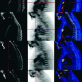
Fig. 26.1
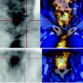
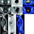
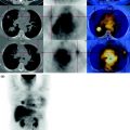
In the MIP image a small right pulmonary nodule with restricted carbohydrate consumption is detected, of metastatic nature. The CT scan shows a right paramedian pelvic mass attributable to the uterus, hypodense, that at the PET scan shows intense cellular activity, with underactive area contained within, which is due to colliquation. The right ovary is highly increased in size, appears as hypodense, with fluid content and shows no pathological glucose metabolism at the PET
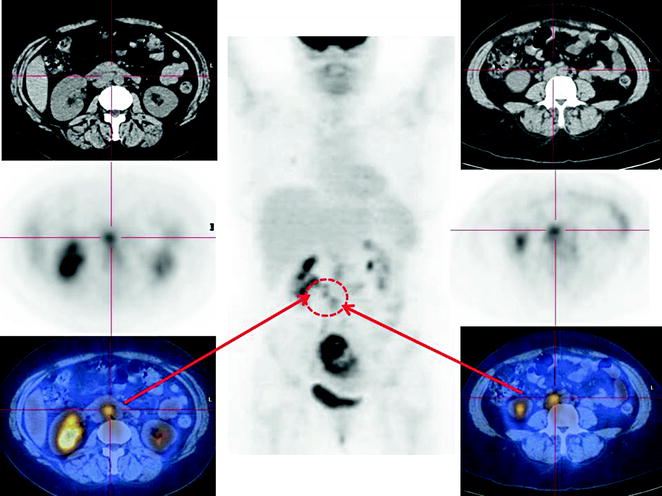
Fig. 26.2


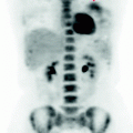
CT-PET shows two lymph nodes increased in size, characterized by high metabolism of glucose, respectively, in the intercavo-aortic and iliac regions. This is associated with a moderate right hydronephrosis secondary to extrinsic nodal compression. Absence of obstruction in place
Stay updated, free articles. Join our Telegram channel

Full access? Get Clinical Tree


