Introduction
Computed tomography (CT) scans have become the mainstay modality for screening patients in the setting of cervical trauma and identifying fractures in the subaxial cervical spine. High-resolution CT imaging, in particular, has become the primary radiologic means for screening patients at most trauma centers, largely replacing radiographs given a superior sensitivity (90%–100%) in identifying traumatic bony injuries. It is therefore critical for spine surgeons and neuroradiologists to understand: (1) normal subaxial cervical anatomy on CT imaging, including commonly used measures of spine relationships and CT-based landmarks for discs and joint spaces; (2) CT findings in the setting of subaxial cervical spine trauma; and (3) CT-based scoring systems for subaxial cervical spine injury.
Subaxial Cervical Spine Measurements and Anatomical Relationships on CT Imaging
Coronal CT Imaging
In coronal CT imaging of the subaxial cervical spine, three of the most important measurements and anatomical relationships to understand include the uncovertebral joints (UVJs), the facet joints, and the disc heights.
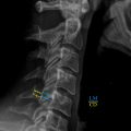
The UVJs are articulations between the uncinate processes and the inferior vertebral bodies ( Fig. 1 ). They are located between the five vertebral bodies of C2–7 (C2–3, C3–4, C4–5, C5–6, and C6–7) and form part of the anterior wall of the vertebral foramen. Functionally, the UVJs provide posterolateral reinforcement of the intervertebral disc, prevent posterior translation of the vertebral bodies, and provide stability to the cervical spine during movement. UVJ measurements can be obtained via CT imaging by measuring at the inferolateral corner of the vertebral body to the corresponding uncinate process of the vertebral body as discussed later. Previous studies have suggested that the mean length of the UVJ on coronal CT imaging in normal anatomical subjects ranged from 1.27 to 1.87 mm, with expected variations between different cervical levels.
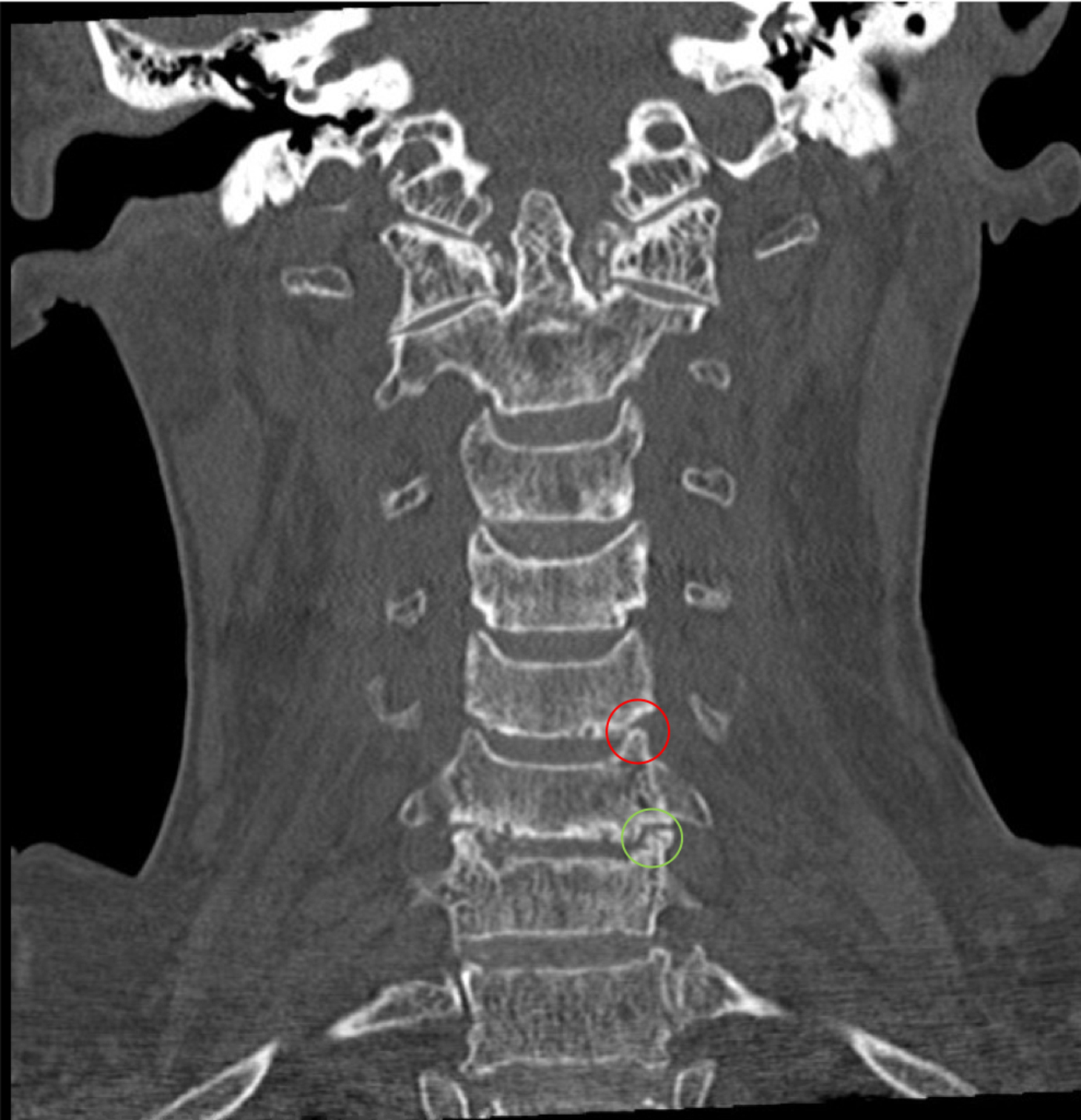
The intervertebral disc height is another important measurement that may be examined in coronal CT imaging of the subaxial cervical spine. The height of the intervertebral discs may be measured on coronal imaging by taking the vertical length of the disc at the point halfway between the left and right UVJ measurements ( Fig. 2 ). The average disc height in the subaxial cervical spine has been reported to a range from 4.62 to 5.03 mm in CT studies conducted on patients without cervical pathologies.

The facet joints represent another important articulation that lends stability to the subaxial cervical spine by holding the adjacent vertebrae together via connections between the superior and inferior articulating facets. In the cervical spine, the superior facets face superiorly and medially, while the inferior facets are oriented inferiorly and laterally. Three different measurements can be taken of the cervical facet joint space on coronal CT imaging, including measurements of the lateral, medial, and midpoint of the joint space. The lateral and medial portions of the facet joint space are measured at the most lateral and most medial edges of the joint from a line drawn perpendicularly to the edges of the bones. The middle of the facet joint is measured as the midpoint between the lateral and medial facet measurements. The mean measurements for the medial, lateral, and midpoint portions of the subaxial cervical facet joints have been reported as 0.50–0.68 mm, 0.48–0.54 mm, and 1.04–1.37 mm, respectively, in normal anatomical subjects.
Sagittal CT Imaging
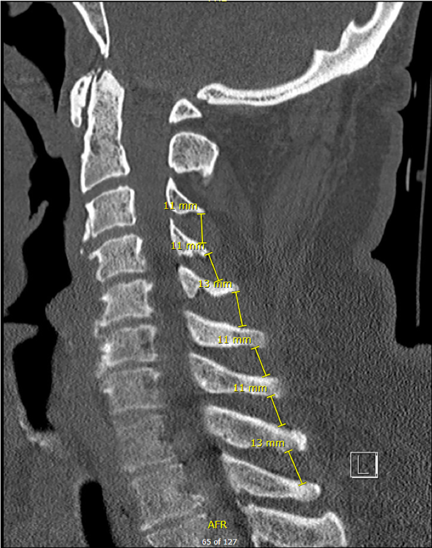
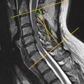
There are several critical bone and joint measurements that one should examine on sagittal CT imaging of the subaxial cervical spine to reflect spinal articulation, alignment, and positioning. These measurements include the interspinous distances (ISDs); vertebral translations; anterior, posterior, and middle facet distances; and anterior, posterior, and middle disc heights.
The ISD represents the distance between the spinous processes of two adjacent spinal segments and may be altered following acute trauma or in degenerative processes in the spinal column. The ISD may be measured by taking the distance between the superior edges of the most posterior portion of adjacent spinous processes on sagittal CT imaging, as demonstrated in Fig. 3 . Previous studies have reported mean ISD values ranging from 13.15 to 15.76 mm in the normal subaxial cervical spine depending on the segments involved.
Vertebral body translation, reflecting shifts in the anterior/posterior positioning of a vertebral body relative to adjacent vertebral bodies, may also be assessed with sagittal CT imaging. This measurement may be obtained by drawing lines parallel to the anterior cortical surfaces of two adjacent vertebral bodies and measuring the distance between these two lines. The typical value for the anterior/posterior vertebral body translation in the sagittal axis of normal patients has been reported to be between 0.09 and 0.58 mm in the subaxial cervical spine.
From sagittal CT imaging, the anterior and posterior portions of the cervical facet joint spaces can easily be visualized and measured using the most anterior and posterior edges of the facets. Similarly, the middle of the facet joint space can be measured at the midpoint of the facet joint between the anterior and posterior measurements. Similarly, one may also determine the percent of facet overlap. Changes or large deviations in the facet overlap may reflect certain pathological states in the facet joints. The percentage of facet overlap may be determined by taking the length of the superior and inferior facet that is covered by each other and dividing it by the total length from the most anterior aspect of the cranial facet to the most posterior aspect of the caudal facet. The mean value for the anterior facet measurement in healthy subjects has been reported to be between 0.83 and 0.92 mm in the subaxial cervical spine, while the posterior facet measurement ranges from 0.56 to 0.76 mm and the middle facet ranges from 0.97 to 1.27 mm.
In a similar manner, the anterior, posterior, and middle portions of the intervertebral discs may be measured on sagittal CT imaging to comprehensively determine disc height in the subaxial cervical spine. The sagittal disc height may be obtained for the anterior and posterior portions of the intervertebral discs, using the anterior and posterior edges of the corresponding vertebral bodies, respectively. The line measuring the disc height is always drawn perpendicularly from the cortical edges of the vertebral bodies that interface with the intervertebral disc. A midpoint measurement of the intervertebral disc height may also be obtained halfway between the anterior and posterior measurements. Previous studies have reported that the mean anterior intervertebral disc height in the subaxial cervical region of healthy subjects has ranged from 2.70 to 3.46 mm. In comparison, the posterior and midpoint disc heights have ranged from 1.99 to 2.45 mm and from 4.66 to 5.24 mm, respectively.
Subaxial Cervical Spine CT Findings in Trauma
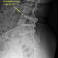
Approximately 75% of cervical spine dislocations and 65% of cervical spine fractures occur in the subaxial region. There are several types of subaxial cervical injuries that CT imaging is well-suited to assess. These include compression and burst fractures, distraction injuries, hyperflexion and hyperextension injuries, and translational or rotational injuries.
Compression and Burst Injuries
Compression and burst injuries present with a loss of height in the fractured vertebral body and do not show any signs of concomitant translational or distraction injury. On CT scans, a fracture line or signs of endplate disruption are often observed in either sagittal or coronal sections ( Fig. 4 ). Compression injuries also encompass fractures of the lateral mass, laminar, facet, and spinous processes that are only minimally displaced and lack any signs of distraction.

Related posts:
 Full-Length Spine CT and MRI in Daily Practice
 Full-Length Spine—Plain Radiographs
 Upper Cervical Spine: Computed Tomography
 Clinical Correlations to Specific Phenotypes and Measurements With Classification Systems
 Magnetic Resonance Imaging Techniques for the Evaluation of the Subaxial Cervical Spine
 Lumbosacral Spine Plain Radiographs
Stay updated, free articles. Join our Telegram channel

Full access? Get Clinical Tree


