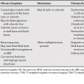
129 The differential diagnosis of subchondral bone marrow edema is broad, but can be narrowed with clinical history and associated findings. Potential trauma or infection can be evaluated by history and a tumor should have an associated focal lesion. After these have been excluded, a cartilage defect or diffuse cartilage thinning associated with the abnormal bone marrow signal should be sought for as osteoarthritis is a common cause of abnormal subchondral signal. Tendon abnormalities (e.g., epicondylitis) often result in abnormal signal in the subjacent bone, although this is usually not subchondral in location. If a subchondral fracture is seen the differential diagnosis in the knee is subchondral insufficiency fracture versus spontaneous osteonecrosis of the knee (SONK) and a subchondral insufficiency fracture versus subchondral fracture associated with osteonecrosis in the hip. Several imaging findings are helpful in differentiating these entities.
Subchondral Bone Marrow Edema
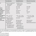
Subchondral Insufficiency Fracture versus Osteonecrosis with Subchondral Fracture
Related posts:

Stay updated, free articles. Join our Telegram channel

Full access? Get Clinical Tree


