and Zdeněk Fryšák1
(1)
Department of Internal Medicine III – Nephrology, Rheumatology and Endocrinology, Faculty of Medicine and Dentistry, Palacky University Olomouc and University Hospital Olomouc, Olomouc, Czech Republic
Keywords
Substernal goiterRetrosternal goiterUltrasoundComputed tomography11.1 Essential Facts
According to the most commonly used definition, the substernal goiter (SSG) or retrosternal goiter (RSG) is one with more than 50% of its mass lying below the thoracic inlet [1].
Prevalence of SSG (depending of definition) ranges from 2–19% among all patients with a goiter [2].
Intrathoracic goiters account for 3.1–5.8% of all mediastinal masses [3].
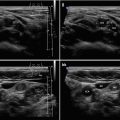
Primary SSG (Fig. 11.1aa) (an ectopic thyroid tissue detached from a cervical thyroid mass, receiving blood supply from mediastinal vessel) is very rare (1%). Secondary SSG (Fig. 11.2aa) is more common as a part of multinodular goiter, with its portion extending retrosternally [4].
Patients are generally in the fifth decade of life, and women predominate.
Many patients experience dysphagia (52%), shortness of breath (52%), voice issues (11%), and chest pressure (12%) [5].
In a large analysis of 80 patients with SSG, postoperative histology revealed multinodular goiter in 51%, follicular adenoma in 35%, Hashimoto’s thyroiditis in 5%, and occult papillary carcinoma 1.6% [6].
Some prospective studies document the incidence of carcinoma development in SSG at 1.3–3.7 new cases per 1000 patients [7].
The incidence of thyroid cancer in SSG is not higher than the incidence of cancer in cervical goiters [8].
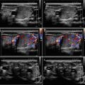
Fig. 11.1
(aa) A 50-year-old woman with primary substernal multinodular goiter (SSG), size 55 × 53 × 35 mm and volume 53 mL (entirely visible and measured by US) on the right side. US overall view of atrophic thyroid gland in thyroid bed: homogeneous structure; isoechoic; Tvol 5 mL, RL 3 mL, and LL 2 mL; transverse. Note: pictogram—thick arrow indicates location of SSG (not shown)—caudally from the current probe position. (bb) Detail of atrophic RL and upper pole of SSG: atrophic RL—homogeneous structure; isoechoic; upper pole of SSG—coarse structure; hyperechoic; clear 15 mm space between low pole of the RL and upper pole of SSG; longitudinal, depth of penetration 5 cm. (cc) Detail of space between atrophic RL and upper pole of SSG: clear 15 mm space between low pole of the RL and upper pole of SSG; longitudinal. (dd) Detail of separated SSG: solid; inhomogeneous structure; hyperechoic; probe inclined retrosternally; transverse. (ee) Detail of separated SSG: solid; inhomogeneous structure; hyperechoic; probe inclined retrosternally; longitudinal