The glenohumeral joint provides the greatest range of motion of any joint in the human body. Over the past several decades, histologic studies, biomechanical studies, and improved arthroscopic techniques have contributed to improved knowledge and treatment of glenohumeral joint abnormalities. Continuing advances in magnetic resonance technology have allowed for improved noninvasive visualization of the stabilizers of the shoulder. This article reviews the concept of glenohumeral joint microinstability and its relationship with superior labrum anterior and posterior (SLAP) lesions, reviews the role of the labrum as a stabilizer of the shoulder, and focuses on the diagnosis and classification of SLAP lesions.
The glenohumeral joint provides the greatest range of motion of any joint in the human body. To achieve this mobility, complex interactions between several static and dynamic stabilizers must occur. Any injury that disturbs these interactions can lead to altered movement between the humeral head and the glenoid.
Over the past several decades, histologic studies, in vivo and in vitro biomechanical studies, and improved arthroscopic techniques have contributed to improved knowledge and treatment of abnormalities of the glenohumeral joint, including the labrum. In particular, continuing advances in magnetic resonance (MR) technology have allowed for improved noninvasive visualization of the stabilizers of the shoulder. In this article, the authors review the concept of glenohumeral joint microinstability and its relationship with superior labrum anterior and posterior (SLAP) lesions, review the role of the labrum as a stabilizer of the shoulder, and focus on the diagnosis and classification of SLAP lesions.
Microinstability of the glenohumeral joint
Definition and Pathoetiology
The concept of microinstability is challenging from a clinical standpoint as well as in its description in the literature. There is no universally accepted definition. Furthermore, the literature describes microinstability in markedly different patient groups with multiple mechanisms of injury to different structures. At the most basic level, some use the term macroinstability interchangeably with dislocation and define microinstability as any rotational or directional pathologic laxity that leads to abnormal mechanics within the shoulder without frank dislocation. A more specific and generally agreed definition of microinstability has been used to describe the pathology in the superior half of the glenohumeral joint with resultant abnormal translation of the humeral head on the glenoid. This condition leads to subluxation without dislocation because of the limits to translation imposed by the coracoid, acromion, coracoacromial ligament, and rotator cuff. A review of the literature yields 2 viewpoints of microinstability, a clinical classification group and a structural abnormality group, which are not mutually exclusive and demonstrate shared concepts.
In the clinical classification group, microinstability lies between the spectrum of TUBS (Traumatic instability, Unidirectional in nature, with a Bankart lesion typically responding to Surgery) and AMBRII (Atraumatic instability, Multidirectional in nature with Bilateral shoulder findings, which may respond to a Rehabilitation program or require an Inferior capsular shift or Interval closure). Two distinct cohorts have been described classically: young individuals with chronic repetitive microtrauma from overhead motions (also referred to by some as AIOS, Acquired Instability in Overstressed Shoulder) and those without chronic overhead motions.
With regard to overhead athletes, and specifically in the throwing shoulder of baseball pitchers, some have classified internal impingement as microinstability. Although abnormal function in overhead athletes may be a spectrum with some overlap of impingement and instability, most of the literature on this topic delineates the 2 entities. In 1991, Walch and colleagues described internal impingement as an intra-articular impingement that occurs in all shoulders in the abducted externally rotated position. In this position, the undersurface of the posterosuperior rotator cuff may become pinched between the labrum and greater tuberosity. Some consider that this phenomenon of impingement leads to abnormality of the throwing shoulder, whereas others do not. Those that do not consider impingement to be a pathologic entity describe a separate pathologic cascade leading to the dead shoulder of throwing, which involves the SLAP lesion. In this group, repair of the SLAP lesion with physical therapy is usually curative.
Regarding the group of patients who do not use chronic forceful overhead motions, there are 2 additional subsets: those with trauma and those without trauma. The more common subset in this group is traumatic and includes most patients with SLAP lesions as described by Snyder and colleagues (a total of 23 patients, 13 of whom had a compression force to the shoulder and 6 of whom had a sudden pull on the arm). This subgroup would also include most of the patients described by Savoie and colleagues with superior labrum anterior cuff (SLAC) lesions (a total of 40 patients, 6 of whom were in motor vehicle accidents and 15 of whom had traumatic falls onto the shoulder). A much rarer subset of this second group of patients who do not use overhead motions is atraumatic with typical complaints of shoulder pain after a period of inactivity such as pregnancy or immobilization, classified as AMSI (Atraumatic Minor Shoulder Instability). AMSI patients may have static anatomic variants of the middle glenohumeral ligament (MGHL) (absence, hypoplasia, or a Buford complex). In the literature, the term minor instability has been used to encompass both AIOS and AMSI.
Microinstability: Structural Abnormality Group of Literature
From the diagnostic radiologist’s perspective, the structural abnormality class of microinstability is the more useful one. In the literature, this class of microinstability includes patients of varying ages with multiple mechanisms of injury. One article estimates that the incidence of microinstability is 5% in those with shoulder pain. This group shares the etiology of acute trauma or repetitive stress, leading to injury of one or more supporting structures of the superior half of the glenohumeral joint, including injury to the labrum, MGHL, rotator interval structures, and rotator cuff. Injuries to any of these supporting structures can lead to predictable pathologic translation of the humeral head relative to the glenoid.
Labral injuries constitute the largest portion of this group (discussed in detail in the following sections). The MGHL has been shown to be an important secondary restraint to both inferior and anterior translation ( Fig. 1 ). Savoie and colleagues reported on isolated MGHL avulsions in 33 patients with anterior instability, who were treated with improvement in pain and function.

The rotator interval includes the superior glenohumeral ligament (SGHL), coracohumeral ligament, glenohumeral joint capsule, and biceps tendon, all of which contribute to stability. The SGHL has been shown to be an important secondary restraint to anterior and superior translation of the humeral head in shoulder flexion and lesser degrees of abduction. Injury to the SGHL can lead to anterosuperior instability ( Fig. 2 ) and secondary impingement of the rotator cuff tendon. With regard to this specific association, the term SLAC was introduced by Savoie and colleagues to describe anterosuperior labral lesions, SGHL avulsion or injury, and anterior cuff pathology. Studies have shown that injury to the rotator interval or other containing structures can result in increased posterior and inferior translation with the arm in neutral position and increased anterior translation with the arm flexed 60°.

Labral anatomy and biomechanical considerations
The glenoid labrum is a vascularized ring of fibrous tissue that surrounds the bony glenoid. The labrum serves numerous functions, including providing nutrition to the glenoid cavity and maintaining joint lubrication. Biomechanically the labrum stabilizes the joint by deepening the glenoid fossa and increasing articular surface area by approximately one-third. The labrum also acts to protect the chondral surface from compression and sheer damage, and serves as an attachment for capsuloligamentous structures, biceps, and triceps. In addition, it acts as a valve that maintains negative intra-articular pressure and stability.
For localization purposes, the labrum can be divided into 4 (superior, anterior, inferior, posterior or anterosuperior, anteroinferior, posteroinferior, posterosuperior) or more circular sectors. Alternatively, the labrum can be divided into a clock face with the most common designation using 12 o’clock to represent superior and 3 o’clock to represent anterior. The initial descriptions of SLAP extended between 10 and 2 o’clock. However, designation of the precise 12 o’clock location can be difficult depending on which structure is used. A study on the biceps anchor showed that if the supraglenoid tubercle is used to represent 12 o’clock, the biceps anchor was situated at 12 o’clock 44% of the time and posterior to this 51% of the time (11 o’clock with reference to the supraglenoid tubercle).
From a functional point of view, the labrum, biceps tendon, and glenohumeral ligaments can be considered as a single functional unit. Huber and Putz performed macroscopic and microscopic analyses, which showed bundles of parallel collagen fibers that run around the entire circumference of the glenoid and into the surrounding structures, termed the periarticular fiber system (PAFS). In their study, the bulk of the substance at the posterosuperior aspect of the labrum actually consisted of extended periarticular fiber bundles from the biceps tendon. Huber and Putz concluded that the labrum is not to be regarded as an isolated structure. These results were most recently confirmed by Arai and colleagues, who additionally described a histologically distinct structure that they termed the sheetlike structure, coursing from the glenoid neck and labrum with extension into the rotator interval. This distinct membranous structure courses differently from the SGHL and contains numerous elastic fibers in a crimp pattern, suggesting that it is subject to constant tensile stress.
In some individuals, however, the PAFS demonstrates variability. Pal and colleagues found that in approximately 25% of their 24 specimens, the long head of the biceps tendon originated principally from the supraglenoid tubercle, not the labrum. Others have reported that the long head of the biceps tendon was seen to originate from the undersurface of the rotator cuff or even extra-articularly without attachment to either the superior labrum or supraglenoid tubercle. The glenohumeral ligaments also demonstrate considerable variability. The MGHL has been described to be the most variable of the glenohumeral ligaments, being present between 70% and 85% of the time in anatomic and MR studies, with a variable origin. Less variable is the SGHL, the anatomy of which was more recently elucidated by Kask and colleagues as being composed of oblique and direct fibers. The direct fibers were seen to invariably originate from the glenoid labrum whereas the oblique fibers originated from the supraglenoid tubercle except for those few specimens with absence of the MGHL, with both oblique and direct fibers originating from the labrum in these cases. Patient variability likely influences predisposition to certain patterns of injury, given an identical mechanism.
Labral variants
In addition to variability of the perilabral structures, there is a wide range of variability of the labrum itself. This range includes labral morphology and signal intensity as well as common anatomic variations that have been described in the superior and anterosuperior glenoid region, including the sublabral sulcus, sublabral foramen or hole, and the Buford complex. Multiple variations can occur in the same individual, for instance a sublabral foramen with a cordlike MGHL.
Labral morphology varies depending on the region. The inferior half of the labrum is usually rounded and firmly continuous with the articular cartilage, with detachment considered an abnormal finding. By contrast, the superior and anterosuperior labrum can be rounded or meniscal in shape (roughly triangular) and can be normally mobile. The superior labrum can be categorized into a triangular type, bumper type, or meniscoid type depending on shape and extent onto the articular surface. In one study of 52 shoulders in young asymptomatic patients, there was extensive variability in morphology regarding the dominant features of the anterior and posterior labra, respectively: triangular (45%, 73%), round (19%, 12%), cleaved (15%, 0%), notched (8%, 0%), flat (7%, 6%), and absent (6%, 8%). These studies show that labral morphology is widely varied, more so anteriorly than posteriorly and much more so superiorly than inferiorly.
Increased signal intensity in the labrum may be caused by a magic angle phenomenon, particularly in the posterosuperior aspect. The magic angle phenomenon is a well-recognized nuclear MR artifact seen in clinical imaging of tissues containing well-ordered collagen fibers. In these tissues, dipolar interactions between water hydrogen protons occur along the long axis of the collagen fibers. This restricted motion causes rapid T2 relaxation and essentially no signal on typical spin-echo images. However, when the long axis of collagen bundles is oriented approximately 55° to the main magnetic field (B0), dipolar interactions are minimized and T2 relaxation time lengthens approximately 100-fold, causing increased signal. This artifact can be avoided by confirming abnormal signal intensity on longer echo-time images. However, even in regions of the labrum not typically demonstrating a magic angle, increased signal intensity does not necessarily indicate a tear. Some reports indicate that this can be seen as a normal finding or be caused by degeneration.
Numerous anatomic variations occur in the superior and anterosuperior portions of the glenoid region, which poses a diagnostic challenge because it is also a common location for abnormalities. In 1992, Cooper and colleagues first described the sublabral recess, which occurs beneath the superior labrum and long head of the biceps origin and is covered by a synovial lining. Only at this region does the hyaline articular cartilage of the glenoid extend over the edge of the glenoid rim and onto the superior neck of the scapula. Multiple studies using cadavers and imaging have found the incidence of the sublabral recess to be up to 75%. The sublabral recess can extend posterior to the biceps anchor, particularly when the biceps anchor is primarily to the anterior labrum. In the radiologic literature, the appearance of this recess has been designated as the single Oreo cookie sign, whereby on the coronal images the recess (white cream) is interposed between the labrum and glenoid (black cookies). Numerous studies have shown that the most common cause of interpretation errors lies in the difficulty of differentiation of SLAP type II lesions from sublabral recesses.
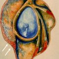
Although not without exceptions, several signs have been described to help in delineating sublabral recesses from SLAP lesions. Characteristics of the high-signal region interposed between the superior labrum and adjacent glenoid rim that have been shown to support a SLAP lesion include extension laterally as it extends superiorly (in contradistinction to extension medially with superior extension, which would point toward the patient’s head), irregular borders ( Fig. 3 ), width on transverse images greater than 2 mm on conventional MR and 2.5 mm on MR arthrography, an uneven width anterosuperiorly and posterosuperiorly ( Fig. 4 ), and extension posterior to the long head of the biceps tendon. Although all of these signs have some support in the literature, additional reports have indicated low to moderate accuracy with some, particularly with increased width between the glenoid and labrum and posterior extension beyond the biceps anchor. In cases where the ancillary finding of a perilabral ganglion cyst is present, diagnosis of a labral tear can be made with confidence ( Fig. 5 ).
Nearby, but distinct from the sublabral recess, is the sublabral foramen, located in the superior aspect of the anterior labrum. The sublabral foramen, otherwise referred to as the sublabral hole, refers to the absence of labral attachment in the anterosuperior glenoid quadrant, and has been reported to be seen in approximately 12% of patients. In distinction from the sublabral recess, which is located at the site of attachment of the biceps tendon, a sublabral foramen is anterior to the biceps anchor. The term Buford complex was coined to describe a cordlike MGHL continuous with the superior labrum and an absence of anterosuperior labral tissue, found in up to approximately 7% of patients.
True incidence of these variations in the general population is not known because studies are not based on a population of healthy shoulders in randomly selected individuals. It is also not known whether these variants represent normal development or are acquired. Some studies have shown that these variants are not present in very early development but occur after the third trimester of life. It has been hypothesized that these variants are a consequence of physiologic mobility and are not a sign of instability.
However, numerous studies have demonstrated that certain variant superior and anterosuperior labral anatomy predisposes to abnormality. Rao and colleagues prospectively evaluated 546 patients who underwent arthroscopy, and after multivariate analysis discovered that the presence of a sublabral foramen with or without a cordlike MGHL and those with Buford complexes were significantly associated with labral fraying and SGHL abnormality. In addition, the presence of a sublabral foramen and a non-cordlike MGHL had a higher prevalence of type II SLAP lesions. Other studies have also demonstrated that the Buford complex is highly predisposed toward superior labral lesions. Each of these studies postulates a mechanism for the increased association with pathological conditions; however, there is no consensus.
Regardless, these variations are important because they need to be distinguished from true SLAP lesions, and repair of these anatomic variations can have adverse consequences. For instance, in the original description of the Buford lesion, when the cordlike MGHL was unknowingly shortened by attachment to the glenoid rim, the patient was left with constrained external rotation.
SLAP lesions
Pathogenesis
The most common mechanisms for SLAP include direct compression loads, forceful traction loads to the arm, and repetitive overhead motion. Impaction loading in cadavers has shown that SLAP tears are more frequent with forward falls (shoulder flexion) than with backwards falls (shoulder extension). In another biomechanical study, traction on the biceps tendon reproducibly created a SLAP II lesion, with inferior glenohumeral subluxation facilitating the tear.
With regard to the overhead athlete, several anatomic and biomechanical factors predispose this group to SLAP tears. As already described, some consider that repetitive contact and internal impingement of the labrum with the undersurface of the cuff in the abducted externally rotated position can cause abnormalities. Although the exact mechanism remains to be elucidated, subsequent controlled laboratory studies have shown that SLAP II lesions were seen more frequently in this late cocking phase of throwing. Another study found increased mechanical strain at the posterosuperior labrum in the late cocking phase, which was thought to be due to the anatomic orientation of the long head of the biceps tendon in this position. Injury during the late cocking phase of throwing is not universally accepted, however. In the first published series on superior labral injury in the overhead athlete, Andrews and colleagues postulated a deceleration mechanism for labral injuries as the biceps contract to slow down the rapidly extending elbow in follow-through. In a laboratory study examining the stages of throwing during simulated biceps loading, Yeh and colleagues found that the greatest stress at the superior labrum/biceps tendon interface occurred during the deceleration stage.
In an elegant series on the throwing shoulder, Burkart and colleagues proposed a biomechanical cascade beginning with an acquired tight posteroinferior capsule, leading to a posterosuperior shift in the glenohumeral rotation point, which results in increased clearance of the greater tuberosity over the glenoid during the lack cocking phase. This process causes increased shear forces at the biceps anchor/posterosuperior labral attachment and failure from their attachments via the peel-back mechanism.
As discussed in the section on Labral Variants, it is unclear how labral variants such as the sublabral foramen or the Buford complex predispose to labral tearing. In addition, from the functional point of view, injury may propagate along the PAFS. At the anterior and anterosuperior labrum, the connecting band as described by Huber and Putz consists of fibers connecting the labrum with the superior and inferior glenohumeral ligaments. At the anterosuperior labrum, fibers of the elastic sheetlike structure, which is subject to constant tensile stress, connect with the rotator interval. At the posterosuperior labrum, fibers of the long head of the biceps tendon intermingle with labrum fibers and form a major component of the labrum. However, individual variants of the PAFS and mechanisms for direction of propagation beyond the 4 types of SLAP originally described have not been well described in the literature.
Superior Labral Tears and SLAP Classification
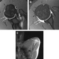
In 1983, Pappas and colleagues presented one of the earliest descriptions of superior labral injury. They proposed the concept that a damaged labrum could cause functional glenohumeral instability and described findings on arthrography. Two years later, Andrews and colleagues reported the first series of superior labral injury in the overhead athlete in 73 baseball pitchers, and described the classic mechanism of labral tearing caused by forces transmitted through the long head of the biceps tendon. In 1990, the term SLAP lesion was introduced to the literature by Snyder and colleagues who described 4 subtypes, occurring with 2 distinct mechanisms of injury: compression force from a fall onto an outstretched arm and traction force caused by a sudden pull on the arm or during overhead sports motion. In type I, the superior labrum is frayed but the peripheral labral edge remains attached to the glenoid ( Fig. 6 ). In type II, the superior labrum and attached biceps tendon are stripped from the glenoid (see Fig. 4 ). In type III, a bucket-handle tear of the superior labrum is present. At least half of the time, SLAP III lesions show the characteristic Cheerio sign, which is a rounded core of soft tissue surrounded by a rim of contrast material ( Fig. 7 ). In type IV, a bucket-handle tear is present with additional extension into the biceps tendon ( Fig. 8 ).
Since 1991, several additional types of SLAP lesions have been described ( Table 1 ). In 1998, Morgan and colleagues described 3 subtypes of SLAP II, type IIA being primarily anterior (see Figs. 3 and 5 ), type IIB being primarily posterior, and type IIC representing combined anterior and posterior tears (see Fig. 4 ). These investigators noted that type IIB and IIC lesions were more frequently observed than SLAP IIA lesions in throwing athletes. Maffet and colleagues described 3 additional types of SLAP after observing other patterns of biceps tendon-superior labrum injury: type V, described as an anterior-inferior Bankart lesion, continues superiorly to include separation of the biceps anchor ( Fig. 9 ); type VI, an unstable flap tear of the superior labrum in addition to biceps tendon separation; and type VII, a superior labrum biceps tendon separation that extends anteriorly beneath the MGHL. In 2004, Nord and colleagues described type VIII, an extended SLAP lesion along the posterior glenoid labrum ( Fig. 10 ); type IX, a circumferential labral tear ( Fig. 11 ); and type X, a SLAP lesion associated with a reverse Bankart lesion ( Fig. 12 ). Another type X lesion exists in the literature, which was described in 2000 as a SLAP lesion that extends into the rotator interval in the form of a lesion of the SGHL, coracohumeral ligament, capsule, or synovium.
Related posts:
 Magnetic Resonance Imaging of Rotator Cuff Disease and External Impingement
 Internal Impingement Syndromes
 The Throwing Shoulder: the Orthopedist Perspective
Magnetic Resonance Imaging of the Pediatric Shoulder
The Throwing Shoulder: the Orthopedist Perspective
Magnetic Resonance Imaging of the Pediatric Shoulder
 Entrapment Neuropathies of the Shoulder
 Anatomic Variants and Pitfalls of the Labrum, Glenoid Cartilage, and Glenohumeral Ligaments
Stay updated, free articles. Join our Telegram channel

Full access? Get Clinical Tree


